
Comprehensive Thesis
1. Introduction
The human circulatory system is one of the most sophisticated biological transportation networks in nature. It consists of the heart, arteries, arterioles, capillaries, venules and veins, all working together to maintain the survival of approximately 37 trillion human cells.
At the centre of this transportation network are the blood capillaries.
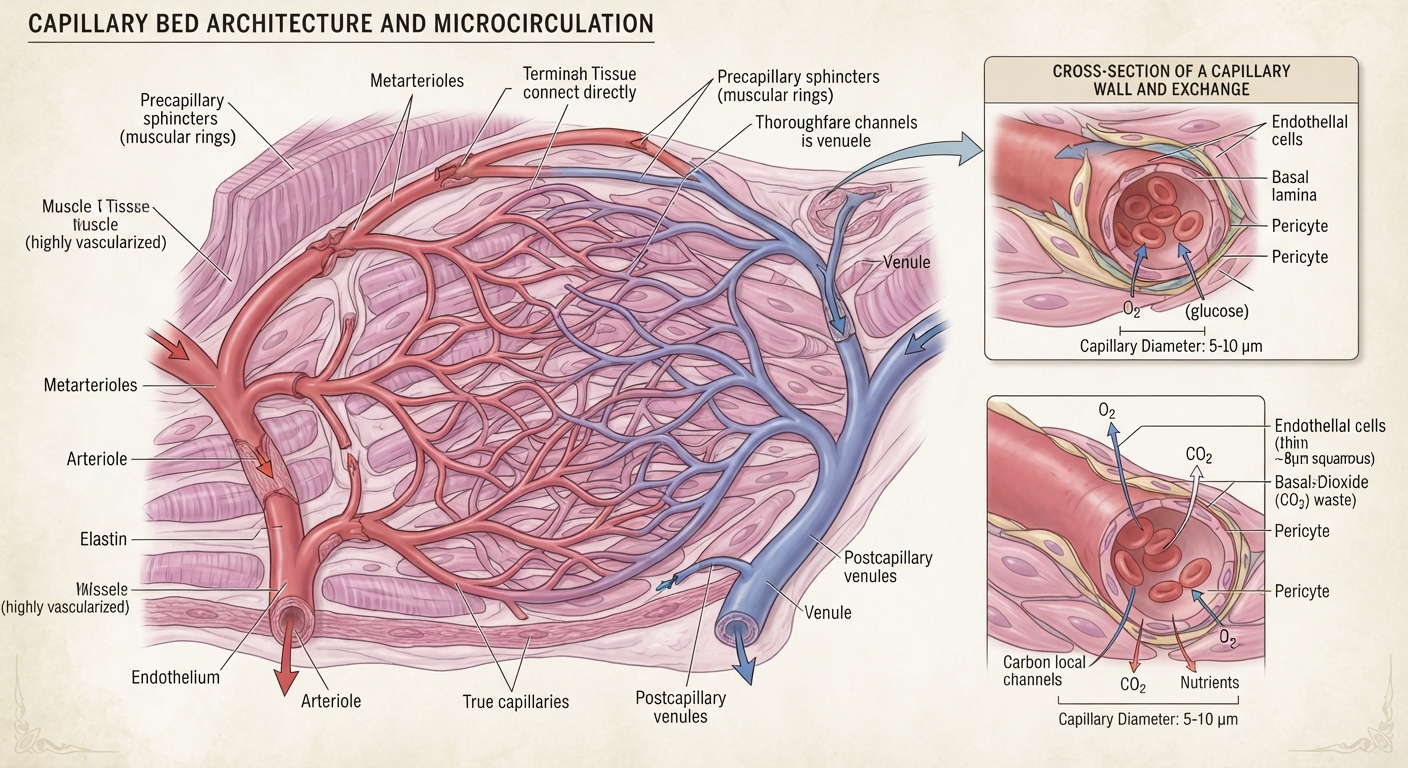
Capillaries are the smallest blood vessels in the human body. Although microscopic, they perform some of the most important functions in human physiology. They connect the arterial circulation to the venous circulation and provide the principal exchange interface between circulating blood and surrounding tissues.
Through their walls, oxygen, nutrients, water, hormones and other substances move between the blood and body tissues. Waste products, including carbon dioxide, also enter the circulation for removal.
An important scientific principle is:
The heart creates the pressure that drives circulation, the arteries distribute blood, the capillaries exchange substances, and the veins return blood to the heart.
Understanding capillary architecture therefore means understanding how the human body transports the materials necessary for cellular survival.
Human A&P exam 2 Flashcards – Easy Notecards
PPT – HISTOLOGI KARDIOVASKULER PowerPoint Presentation, free download – ID:2258884
Pathophysiological and clinical aspects of nonclassical targets of microangiopathy | Oncohema Key
10
2. The complete architecture of the human blood-vessel network
The blood-vessel system is not a collection of independent tubes. It is an interconnected branching network.
Blood follows an organised pathway:
Blood circulation pathway
HEART
Pumps blood into circulation
ARTERIES
Transport blood away from the heart
ARTERIOLES
Control blood distribution into tissues
CAPILLARY NETWORK
Exchange oxygen, nutrients and waste products
VENULES
Collect blood from capillary networks
VEINS
Return blood towards the heart
HEART
Receives returning blood
This sequence represents the general systemic circulation. Pulmonary circulation follows a related pathway through the lungs, where blood releases carbon dioxide and receives oxygen.
3. What exactly is a capillary?
A capillary is a microscopic blood vessel that connects the smallest arterial branches with the smallest venous branches.
Its dimensions are extremely small:
- Typical diameter: approximately 5–10 micrometres, although it varies.
- Wall thickness: generally one endothelial cell layer.
- Structure: a thin endothelial lining supported by a basement membrane.
- Function: exchange between blood and surrounding tissues.
A micrometre (µm) is one-millionth of a metre.
The narrow diameter means that red blood cells often pass through capillaries in single file.
Function Of Capillaries Image – Anatomy System – Human Body Anatomy diagram and chart images
Diffusion in biology explains how minerals leave/enter cells
10
The thin wall is particularly important because it reduces the distance that oxygen and other substances must travel to reach nearby cells.
4. Microscopic anatomy of capillary walls
The capillary wall is structurally simple compared with the walls of larger arteries and veins. However, its components are highly specialised.
OLI – Drawing Structure of different capillaries – English labels | AnatomyTOOL
4.1 Endothelial cells
Endothelial cells form the internal lining of blood vessels.
Their responsibilities include:
- Creating a smooth surface for blood flow.
- Regulating the movement of substances through the vessel wall.
- Participating in blood clotting regulation.
- Controlling inflammatory responses.
- Releasing signals that influence blood-vessel diameter.
- Helping maintain the barrier between blood and surrounding tissues.
Endothelial cells are therefore not merely structural coverings. They are active biological regulators.
4.2 Basement membrane
The basement membrane is a thin supporting layer beneath endothelial cells.
Its functions include:
- Providing structural support.
- Helping maintain vessel integrity.
- Regulating movement of certain molecules.
- Supporting the attachment of surrounding cells.
4.3 Pericytes
Pericytes are specialised cells located around many small blood vessels.
They help with:
- Stabilising capillaries.
- Regulating local blood flow.
- Supporting endothelial cells.
- Participating in tissue repair.
- Maintaining the blood-brain barrier in cooperation with other cells.
4.4 Capillary lumen
The lumen is the internal space through which blood travels.
It accommodates:
- Plasma.
- Red blood cells.
- White blood cells.
- Platelets.
Because the lumen is extremely narrow, blood cells pass through in a tightly organised manner.
5. Three major types of capillaries
The human body has three major structural categories of capillaries. Their differences reflect the functions of the organs they serve.
Blood Vessels – Antranik Kizirian
5.1 Continuous capillaries
Most common type
- Have a continuous endothelial lining.
- Have relatively small gaps between neighbouring cells.
- Permit controlled exchange of water, gases and small molecules.
- Found in muscles, lungs, skin and the nervous system.
- Specialised versions form the blood-brain barrier.
PPT – Blood Vessels PowerPoint Presentation, free download – ID:3026116
5.2 Fenestrated capillaries
- Contain tiny openings called fenestrations.
- Allow relatively rapid exchange of water and dissolved substances.
- Found in the kidneys, small intestine and endocrine glands.
- Important for filtration, nutrient absorption and hormone distribution.
Sinusoid of the Liver Diagram | Quizlet
5.3 Sinusoidal capillaries
- Have wider openings and discontinuous endothelial structures.
- Have a more permeable wall and often an incomplete basement membrane.
- Allow larger molecules and, in certain organs, cells to pass.
- Found in the liver, spleen and bone marrow.
These three types illustrate an important biological principle: different organs require different levels of permeability to perform their specialised functions. NCBI+2
6. The connections between capillaries and major human organs
Capillary networks are distributed throughout almost every living tissue. They are adapted to the metabolic requirements of each organ.
The respiratory system – Clinical Tree
Human Digestive system – Structure, Process
6
6.1 Brain and nervous system
The brain has an extensive network of specialised capillaries.
Their functions include:
- Supplying oxygen to neurons.
- Delivering glucose for energy production.
- Maintaining a controlled chemical environment.
- Helping regulate the blood-brain barrier.
The blood-brain barrier is formed primarily by specialised endothelial cells joined by tight junctions, together with supporting cells and surrounding structures.
It controls which substances can enter brain tissue from the bloodstream.
6.2 Heart muscle
The heart itself requires a continuous supply of oxygen and nutrients.
Its capillaries are located throughout the cardiac muscle, where they support the energy requirements of continuously contracting heart cells.
The coronary arteries supply blood to the heart muscle, which branches into smaller vessels and capillary networks.
6.3 Lungs
The lungs contain millions of tiny air sacs called alveoli.
Each alveolus is surrounded by a dense network of pulmonary capillaries.
File:Cross section of an alveolus and capillaries showing diffusion of gases.svg – Wikimedia Commons
The process works as follows:
- Blood arrives from the right side of the heart through the pulmonary arteries.
- It travels through smaller arteries and arterioles.
- It reaches capillaries surrounding the alveoli.
- Oxygen diffuses from the air inside the alveoli into the blood.
- Carbon dioxide diffuses from the blood into the alveoli.
- Oxygen-rich blood returns to the left side of the heart through pulmonary veins.
This is called pulmonary gas exchange.
6.4 Kidneys
The kidneys contain specialised capillary structures called glomeruli.
These capillary networks filter blood plasma to begin the formation of urine.
30.4.4: Gross Anatomy of the Kidney – Chemistry LibreTexts
The blood pathway is:
Renal artery → smaller arteries → afferent arteriole → glomerular capillaries → efferent arteriole.
An important distinction is that blood does not immediately enter a venule after passing through the glomerulus. It first leaves through the efferent arteriole and enters another capillary network, such as the peritubular capillaries or vasa recta.
These second networks support the exchange of water, nutrients and other substances around the kidney tubules.
6.5 Small intestine
The small intestine contains microscopic projections called villi.
Each villus contains blood capillaries and a central lymphatic vessel called a lacteal.
The blood capillaries absorb many water-soluble nutrients, including glucose and amino acids, and transport them towards the liver through the hepatic portal circulation.
Most absorbed dietary fats enter the lacteals and travel through the lymphatic system before reaching the bloodstream.
6.6 Liver
The liver contains specialised sinusoidal capillaries.
These vessels allow close interaction between blood and liver cells.
Their responsibilities include:
- Processing absorbed nutrients.
- Regulating blood glucose.
- Metabolising various substances.
- Producing important blood proteins.
- Processing substances delivered from the digestive system.
6.7 Skeletal muscles
Muscle tissue has an extensive capillary network.
During physical activity, working muscles require increased oxygen and energy substrates. Their local circulation adjusts to meet these demands.
Capillaries also remove carbon dioxide and other metabolic products.
7. The scientific mechanism of capillary exchange
This is arguably the most important function of the entire capillary system.
Capillary exchange is the movement of substances between blood plasma and the surrounding tissue fluid.
There are four important mechanisms.
CV Physiology | Mechanisms of Capillary Exchange
7.1 Diffusion
Diffusion is the movement of molecules from an area of higher concentration to an area of lower concentration.
Examples:
- Oxygen moves from blood into tissues where oxygen concentration is lower.
- Carbon dioxide moves from tissues into blood.
- Glucose moves from blood into tissues through appropriate transport mechanisms.
7.2 Filtration
Filtration occurs when fluid moves out of capillaries into surrounding tissue spaces due to pressure differences.
This movement helps deliver water and dissolved substances to the tissues.
7.3 Reabsorption
Reabsorption describes fluid moving from tissue spaces back into blood vessels.
The balance of fluid movement depends on several factors, including hydrostatic pressure, plasma protein osmotic pressure and the condition of the endothelial barrier.
An important modern refinement is that sustained reabsorption along the venous end of most ordinary capillaries is not the general rule. Much of the filtered fluid returns through lymphatic vessels.
7.4 Transcytosis
Transcytosis is the movement of substances through endothelial cells using small membrane-bound transport vesicles.
It is particularly important for certain larger molecules that cannot easily cross the endothelial barrier by ordinary diffusion.OpenStax+2
8. The mathematical physics of capillary circulation
Capillary circulation can be understood through several scientific disciplines:
- Physics: pressure, fluid movement and resistance.
- Mathematics: flow calculations and concentration gradients.
- Chemistry: oxygen, carbon dioxide and nutrient exchange.
- Biology: cellular metabolism and tissue survival.
8.1 Blood flow and pressure
Blood moves because of pressure differences.
A simplified relationship is:
\[ Q=\frac{\Delta P}{R} \]
Where:
- \(Q\) = blood flow.
- \(\Delta P\) = pressure difference.
- \(R\) = vascular resistance.
When resistance increases, blood flow tends to decrease if the pressure difference remains constant.
Arterioles are particularly important regulators of resistance in the circulation.
8.2 Poiseuille’s law
For an idealised cylindrical tube with steady laminar flow:
\[ Q=\frac{\pi r^4\Delta P}{8\eta L} \]
Where:
- \(r\) = vessel radius.
- \(\Delta P\) = pressure difference.
- \(\eta\) = fluid viscosity.
- \(L\) = vessel length.
The fourth-power relationship means that changes in vessel radius can have a substantial effect on resistance.
However, actual blood flow is more complicated because blood contains cells, vessels branch, and biological vessels are elastic.
8.3 Why is blood flow slower in capillary networks?
Although individual capillaries are extremely narrow, the body contains enormous numbers of them operating in parallel.
Their combined cross-sectional area is very large.
This reduces average blood velocity and provides more time and surface area for exchange.
The relationship is:
\[ v=\frac{Q}{A} \]
Where:
- \(v\) = average blood velocity.
- \(Q\) = total volumetric blood flow.
- \(A\) = total cross-sectional area.
The larger the combined cross-sectional area, the lower the average velocity for a given flow rate.
This is one of the most important principles in understanding microcirculation.
9. The relationship between blood capillaries and the lymphatic system
One of the most important connections in human anatomy is the relationship between the blood circulatory system and the lymphatic system.
The two systems work together to maintain the correct fluid balance around body cells.
Lymphatic Capillaries: Anatomy and Role in the Lymphatic System – Anatomy Note
9.1 How the connection works
The process follows this sequence:
- Blood enters capillary networks.
- Some fluid moves through the capillary wall into surrounding tissue spaces.
- This fluid becomes part of the interstitial fluid.
- Body cells exchange nutrients, gases and waste products with this fluid.
- Excess interstitial fluid and escaped proteins enter lymphatic capillaries.
- Lymph travels through larger lymphatic vessels.
- Lymph eventually returns to the bloodstream through large veins near the neck.
9.2 What is interstitial fluid?
Interstitial fluid is the fluid occupying the spaces between cells.
It provides the immediate environment through which many substances travel between blood vessels and cells.
Its composition includes:
- Water.
- Dissolved electrolytes.
- Nutrients.
- Hormones and signalling molecules.
- Waste products.
- Proteins in varying concentrations.
9.3 What happens if this connection fails?
If the lymphatic system cannot adequately drain excess tissue fluid, fluid can accumulate in tissues.
This condition is called lymphoedema.
The lymphatic system therefore performs an essential supporting role in maintaining blood volume, tissue fluid balance and immune defence.NCBI+1
10. The role of red blood cells inside capillaries
Red blood cells, also called erythrocytes, are specialised transportation cells.
Their primary responsibility is to carry oxygen using a protein called haemoglobin.
10.1 Red blood cell architecture
A mature human red blood cell has:
- A biconcave disc shape.
- A flexible cell membrane.
- Haemoglobin molecules.
- No nucleus.
- No mitochondria.
Its shape increases its surface-area-to-volume ratio and assists gas exchange.
10.2 Oxygen transportation
In the lungs:
\[ Hb+O_2 \rightleftharpoons HbO_2 \]
Haemoglobin combines reversibly with oxygen.
In tissues, oxygen is released according to local conditions and cellular demand.
The oxygen then diffuses towards cells and their mitochondria.
10.3 Connection to mitochondria
Mitochondria are the principal sites of aerobic ATP production in human cells.
Oxygen is used in the electron transport chain during oxidative phosphorylation.
This produces ATP, the energy-carrying molecule that supports cellular functions.
The complete biological transportation pathway is:
Lungs absorb oxygen
Red blood cells carry oxygen
Capillaries deliver oxygen to tissues
Oxygen enters cells
Mitochondria use oxygen in ATP production
ATP supports cellular activity
This demonstrates why capillary circulation is essential to human energy metabolism.
11. How the body regulates capillary blood flow
The body does not distribute the same quantity of blood to every organ at every moment.
Blood flow changes according to tissue requirements.
For example:
- During exercise, working muscles need increased blood supply.
- After eating, digestive organs increase their activity.
- During temperature regulation, skin circulation changes.
- The brain requires continuous blood circulation to maintain its functions.
Important regulators
| Regulator | Function |
|---|---|
| Arterioles | Control resistance and blood delivery |
| Endothelial cells | Release substances regulating vessel behaviour |
| Nitric oxide | Helps relax vascular smooth muscle |
| Sympathetic nervous system | Influences blood-vessel diameter |
| Local metabolites | Help match blood supply to tissue activity |
| Pericytes | Support and help regulate microvascular function |
A particularly important distinction is that capillaries themselves generally lack the continuous smooth-muscle layer found in arterioles. Their blood supply is regulated mainly through upstream vessels and local microvascular mechanisms. Physiological Reviews+1
12. The architecture of capillary connections in different body systems
The following table summarises the major anatomical relationships.
| Body system | Capillary connection | Main responsibility |
|---|---|---|
| Nervous system | Brain microcirculation | Oxygen and nutrient delivery |
| Respiratory | Pulmonary capillaries | Gas exchange |
| Digestive | Intestinal villus capillaries | Nutrient absorption |
| Cardiovascular | Coronary capillaries | Heart muscle nourishment |
| Renal | Glomerular and peritubular networks | Filtration and reabsorption |
| Muscular | Skeletal muscle capillaries | Energy and oxygen supply |
| Endocrine | Glandular capillaries | Hormone exchange |
| Hepatic | Liver sinusoids | Nutrient processing |
| Immune and blood-forming | Bone marrow sinusoids | Blood-cell movement |
| Skin | Dermal capillaries | Tissue nourishment and temperature regulation |
13. Capillary health and the importance of maintaining the microcirculation
Capillary function is influenced by the overall condition of the cardiovascular system.
Factors that can affect the microcirculation include:
- High blood pressure.
- Diabetes and prolonged elevated blood glucose.
- Smoking exposure.
- Inflammation.
- Reduced oxygen delivery.
- Blood-vessel damage.
- Abnormal blood clotting.
Some examples of diseases involving small blood vessels include diabetic microvascular disease and certain forms of retinal vascular disease.
The retina is particularly interesting because its capillaries have specialised barriers that help maintain the delicate environment needed for vision.
Why this matters
A person can have large arteries that transport blood while still experiencing problems in the smaller vessels responsible for delivering that blood to tissues.
This is why medical science examines the entire vascular network rather than concentrating exclusively on the heart and major arteries.
14. The relationship between capillary architecture and modern scientific technology
Capillary anatomy has become an important research area in modern medicine, biotechnology and engineering.
Quantification of Diabetes-induced Adherent Leukocytes in Retinal Vasculature
MIT’s new magnetic mixer creates the most uniform 3D-printed tissues
12
Several modern technologies build on knowledge of capillary architecture.
1. Medical microscopy
Advanced microscopy allows researchers to examine endothelial cells, blood flow and microscopic vessel structures.
2. Organ-on-a-chip technology
Researchers construct small laboratory systems that reproduce aspects of human tissue and microvascular environments.
3. Tissue engineering
Scientists investigate ways to develop blood-vessel networks that can supply engineered tissues with oxygen and nutrients.
4. Artificial intelligence in medical imaging
AI can assist researchers and medical professionals in analysing vascular images, identifying patterns and measuring changes in small blood vessels.
5. Regenerative medicine
Understanding how capillaries grow and connect is important for investigating tissue repair and the development of new vascular networks.
These technologies demonstrate how microscopic human anatomy connects with modern engineering and computational science.
15. The complete microscopic connection: from blood to individual human cells
The most remarkable feature of capillary architecture is its proximity to individual cells.
Capillaries form networks throughout tissues, bringing blood close to the cells that depend on it.
The following sequence summarises the entire biological relationship:
STING-associated vasculopathy with onset in infancy: MedlinePlus Genetics
- The heart generates the pressure that drives blood circulation.
- Arteries distribute blood towards organs.
- Arterioles regulate blood delivery.
- Capillaries bring blood close to individual cells.
- Oxygen and nutrients move into surrounding tissues.
- Cells use nutrients and oxygen to maintain their activities.
- Carbon dioxide and other waste products enter the surrounding fluid and circulation.
- Venules collect blood from capillary networks.
- Veins return blood towards the heart.
- The lymphatic system returns excess interstitial fluid and proteins to the bloodstream.
This is a continuous biological transportation and exchange system.
16. Important scientific measurements
| Anatomical characteristic | Approximate information |
|---|---|
| Capillary diameter | 5–10 µm is a common range |
| Red blood cell diameter | Approximately 7–8 µm |
| Capillary wall | One endothelial cell layer with supporting structures |
| Blood-flow velocity | Relatively slow compared with larger arteries |
| Capillary distribution | Throughout almost all vascularised tissues |
| Main exchange mechanism | Diffusion, filtration and selective transport |
| Main supporting structure | Basement membrane |
| Supporting cells | Pericytes in many capillary beds |
These are general reference values. Actual measurements differ between organs and physiological conditions. NCBI+2
17. Final scientific conclusion
The capillary network represents one of the greatest examples of biological engineering.
Its architecture solves an extraordinary transportation problem: how to deliver oxygen, nutrients, hormones and other essential materials to an enormous number of microscopic living cells while collecting metabolic waste.
The heart cannot directly deliver oxygen to every individual cell. Instead, it relies on a hierarchical vascular architecture consisting of arteries, arterioles, capillaries, venules and veins.
Three principles summarise its importance:
- Structural principle: Extremely thin endothelial walls and extensive branching networks provide an effective exchange interface.
- Transportation principle: Blood flow connects the lungs, heart and organs through an integrated circulation.
- Cellular survival principle: Capillary exchange supports the chemical reactions, energy production and maintenance of living tissues.
Ultimately, the capillary system is not simply a collection of microscopic blood vessels. It is the essential exchange infrastructure connecting the cardiovascular system with cellular life.
18. Recommended scientific reference library
For further university-level study, these are useful sources:
- OpenStax Anatomy and Physiology 2e — Blood vessel structure and function.
- NCBI Bookshelf — Histology of capillaries.
- OpenStax — Capillary exchange.
- NCBI Bookshelf — Capillary fluid exchange.
- American Journal of Physiology — Capillary communication and regulation of blood flow.
An important final distinction: The heart is the central pumping organ, but the capillary network is where the circulation performs its essential exchange with living tissues. Understanding both structures together provides a much deeper understanding of human anatomy and physiology.





Be First to Comment