
COMPREHENSIVE SCIENTIFIC THESIS
A detailed scientific tutorial in cellular biology, molecular biology, genetics, biochemistry, human anatomy and ageing science
Chapter 1. Introduction: What are zombie cells?
The term zombie cells is an informal scientific expression used to describe senescent cells in the human body.
These are living cells that have permanently or persistently stopped dividing but have not died.
They remain metabolically active, meaning they continue performing certain biochemical activities. However, their behaviour changes considerably.
Under normal circumstances, cells undergo a life cycle involving:
- Growth and development.
- Normal cellular activity.
- Cell division and multiplication.
- Repair and maintenance.
- Programmed cell death when they are no longer needed.
Senescent cells follow a different biological pathway.
They stop dividing, but they remain inside body tissues. Some release chemical signals that influence neighbouring cells, the immune system and the surrounding tissue environment.
Cellular senescence. Normal cell and senescence cell. Zombie cells. Hallmarks of ageing.
Cellular Senescence: Molecular Targets, Biomarkers, and Senolytic Drugs
“Bone-SASP” in Skeletal Aging | Calcified Tissue International | Springer Nature Link
12
The central scientific question is:
Why does the human body retain cells that have stopped dividing, and how can these cells simultaneously protect the body and contribute to ageing and disease?
The answer involves an important biological principle: cellular senescence is both a protective mechanism and a potential source of tissue dysfunction.
Chapter 2. The molecular architecture of a zombie cell
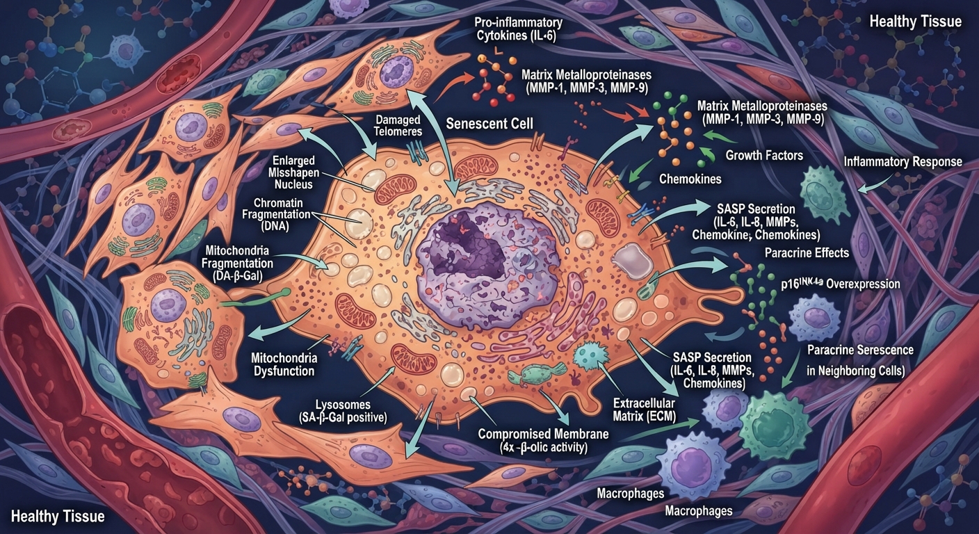
A senescent cell is not an entirely different type of cell. It is an ordinary human cell that has entered a particular biological state.
Its internal architecture contains the same fundamental cellular structures found in other human cells, but several structures and biochemical pathways change.
Cellular Senescence: Molecular Targets, Biomarkers, and Senolytic Drugs
2.1 Main components and their functions
| Cellular component | Function in senescent cells |
|---|---|
| Cell membrane | Controls communication and exchange with surroundings |
| Nucleus | Contains DNA and regulates gene activity |
| Chromosomes | Carry genetic information |
| Mitochondria | Produce energy and influence cellular signalling |
| Ribosomes | Manufacture proteins |
| Endoplasmic reticulum | Processes proteins and lipids |
| Golgi apparatus | Modifies and distributes proteins |
| Lysosomes | Break down and recycle cellular materials |
| Cytoplasm | Contains biochemical machinery |
| Nuclear envelope | Separates and regulates the nuclear environment |
2.2 The nucleus: The control centre
The nucleus contains the cell’s genetic information in the form of DNA.
In senescent cells, the nucleus undergoes several changes:
- DNA damage may accumulate.
- Chromatin organisation changes.
- Certain genes become more active while others become less active.
- Genes associated with cell division are suppressed.
- Genes involved in inflammation and cellular stress responses may become more active.
One important characteristic is the formation of specialised chromatin regions associated with senescence.
These changes help maintain the cell in a non-dividing state.
2.3 Mitochondria: The energy architecture
Mitochondria are particularly important in cellular senescence.
They:
- Produce ATP, the principal energy-carrying molecule.
- Regulate cellular metabolism.
- Participate in programmed cell death.
- Influence cellular stress responses.
- Communicate with other cellular structures.
In senescent cells, mitochondrial function can change.
Some mitochondria become less efficient, while others produce increased levels of reactive oxygen species (ROS).
This can influence inflammatory signalling and reinforce the senescent state.
2.4 Lysosomes: The recycling architecture
Senescent cells frequently develop increased lysosomal activity and enlarged lysosomal compartments.
One important research marker is an enzyme called senescence-associated beta-galactosidase (SA-β-gal).
Its activity is commonly used in laboratory research to help identify senescent cells.
However, it is not sufficient on its own to establish that a cell is senescent.
Chapter 3. How do normal cells become zombie cells?
Cellular senescence does not happen randomly. It is usually triggered by biological stress, genetic damage or changes in the cellular environment.
There are several major pathways.
Figure 1 — The multifaceted inducers of cellular senescence | Aging
3.1 DNA damage
DNA is the instruction manual of the human cell.
It contains genetic instructions for:
- Producing proteins.
- Regulating cellular activities.
- Repairing damaged structures.
- Controlling cell division.
- Maintaining normal tissue function.
DNA can experience damage from normal metabolic processes, environmental factors and errors during DNA replication.
When the damage becomes serious, the cell activates its DNA damage response.
This involves specialised proteins that detect damage, communicate its presence and coordinate repair or other protective responses.
If the damage cannot be safely repaired, the cell may enter senescence.
3.2 Telomere shortening
Telomeres are protective DNA-protein structures located at the ends of chromosomes.
Their function is to protect chromosome ends and prevent them from being incorrectly recognised as broken DNA.
In many dividing human cells, telomeres become shorter after successive divisions.
Eventually, critically short or dysfunctional telomeres can activate cellular damage responses.
The result may be cellular senescence.
This is one reason cellular senescence is associated with biological ageing.
However, not all senescent cells develop because of telomere shortening.
3.3 Oxidative stress
Oxidative stress occurs when the production of reactive oxygen species exceeds the capacity of antioxidant and repair systems to maintain cellular balance.
Reactive oxygen species are chemically reactive molecules produced during normal metabolism and other biological processes.
In controlled amounts, they also participate in cellular signalling.
Excessive oxidative stress can affect:
- DNA.
- Proteins.
- Cell membranes.
- Mitochondria.
- Cellular metabolic pathways.
Severe or persistent damage may contribute to senescence.
3.4 Oncogene activation
An oncogene is a gene that can promote excessive cellular growth when improperly activated.
For example, abnormal activation of growth-promoting pathways can cause a cell to begin dividing excessively.
Interestingly, the cell has protective mechanisms that can detect this abnormal activity.
It may activate senescence to prevent the potentially dangerous cell from multiplying.
This is an important natural defence against cancer.
3.5 Mitochondrial dysfunction
Mitochondria influence cellular energy production and stress signalling.
When their function becomes disrupted, they may contribute to:
- Altered energy metabolism.
- Increased oxidative stress.
- Inflammatory signalling.
- Changes in gene expression.
- Maintenance of cellular senescence.
These five mechanisms demonstrate that senescence is a coordinated biological response rather than simply a cell becoming old.
Chapter 4. The genetic control system: p53, p21, p16 and RB
This is one of the most important chapters for understanding the molecular architecture of zombie cells.
The human body has sophisticated molecular control systems that regulate when cells are permitted to divide.
Four important components are:
JCI – Telomeres, stem cells, senescence, and cancer
4.1 The p53 protein: The genetic guardian
The TP53 gene provides instructions for producing the p53 protein.
Its responsibilities include:
- Monitoring cellular stress.
- Responding to DNA damage.
- Regulating DNA repair.
- Controlling cell-cycle progression.
- Helping initiate senescence or programmed cell death when necessary.
When serious damage is detected, p53 can activate p21.
4.2 The p21 protein: The cell-division regulator
The CDKN1A gene produces the p21 protein.
Its primary role in this pathway is to inhibit particular cyclin-dependent kinases (CDKs).
These kinases are enzymes that help control the cell cycle.
When p21 inhibits their activity, the cell can be prevented from progressing through the division cycle.
4.3 The p16 protein: Another protective regulator
The CDKN2A gene produces p16, also known as p16INK4a.
It inhibits CDK4 and CDK6.
These proteins normally participate in controlling progression through the G1 phase of the cell cycle.
When p16 activity increases, the cell can enter a persistent growth-arrested state.
4.4 The RB protein: The division checkpoint
The RB protein is produced by the RB1 gene.
It regulates E2F transcription factors, which control genes necessary for DNA replication and cell-cycle progression.
When RB is in its active growth-suppressing state, it restricts E2F activity.
This helps prevent the cell from progressing towards division.
The complete molecular relationship
Cellular damage or abnormal growth signals
Pathway A
p53 activation
↓
p21 production
Pathway B
p16 activation
↓
CDK4/6 inhibition
RB-mediated cell-cycle suppression
Protective outcome
Persistent cellular growth arrest
The p53-p21 and p16-RB pathways are closely related, but they are not identical. They can operate together or independently, depending on the type of stress and cell.
The fundamental purpose is to prevent damaged or potentially dangerous cells from continuing to divide.
Chapter 5. The SASP system: How zombie cells communicate with neighbouring cells
One of the most fascinating discoveries in cellular biology is that senescent cells can remain highly active in communication.
Scientists call this the:
Senescence-Associated Secretory Phenotype (SASP).
SASP refers to the collection of biological substances released by senescent cells into their surrounding environment.
Senolytic Interventions for Type 2 Diabetes: Current Evidence and Future Directions | MDPI
5.1 What does SASP release?
| Biological substance | Main function |
|---|---|
| Interleukins | Regulate immune and inflammatory responses |
| Cytokines | Coordinate communication between cells |
| Chemokines | Attract particular immune cells |
| Growth factors | Influence cell growth and repair |
| Proteases | Break down or remodel proteins |
| Matrix metalloproteinases | Remodel the extracellular matrix |
| TGF-β | Regulates tissue repair, inflammation and fibrosis |
5.2 How does communication happen?
Imagine a senescent cell located in human tissue.
It releases chemical signals into the surrounding environment.
These signals can communicate with:
- Nearby normal cells.
- Immune-system cells.
- Fibroblasts.
- Stem and progenitor cells.
- Blood-vessel-associated cells.
Some signals help coordinate tissue repair.
Other signals, particularly when continuously released, may create chronic inflammation.
5.3 Paracrine senescence
An especially important phenomenon is called paracrine senescence.
This happens when chemical signals released by an existing senescent cell encourage neighbouring cells to enter a senescent state.
This can influence the number and distribution of senescent cells within a tissue.
However, it does not mean every neighbouring cell automatically becomes senescent.
The outcome depends on the cell type, signals, biological environment and duration of exposure.
5.4 Why is this important?
SASP illustrates a major principle of human biology:
A cell does not operate independently of the rest of the body.
Cells function within interconnected communication networks.
A change in one cell can influence the behaviour of surrounding cells and, under certain conditions, more distant tissues.
Chapter 6. The relationship between zombie cells and the human immune system
The immune system is one of the body’s important mechanisms for controlling senescent cells.
It can recognise and remove some senescent cells through immune surveillance.
Frontiers | Senescence, NK cells, and cancer: navigating the crossroads of aging and disease
6.1 Natural killer cells
Natural killer (NK) cells are specialised immune cells.
They recognise certain abnormal or stressed cells and can eliminate them.
They participate in the surveillance of senescent cells.
6.2 Macrophages
Macrophages are specialised immune cells that engulf and process unwanted cellular material.
This process is called phagocytosis.
Macrophages can participate in removing senescent cells and coordinating tissue repair.
6.3 T lymphocytes
Certain T lymphocytes help identify abnormal cells and coordinate immune responses.
They can contribute to the recognition and elimination of senescent cells.
6.4 Neutrophils
Neutrophils are important immune cells involved in inflammation and tissue responses.
They can participate in the removal of senescent cells in particular biological contexts.
6.5 What happens when immune clearance becomes less effective?
As the human body ages, immune surveillance can become less effective in certain circumstances.
Some senescent cells may persist longer than they otherwise would.
Their accumulated secretions can contribute to an unfavourable tissue environment.
This can create a biological cycle:
Cellular damage
Senescence develops
Senescent cells release SASP signals
Inflammation and tissue changes
Immune clearance may become less effective
Potential accumulation of senescent cells
This relationship is a major area of research into ageing and age-associated diseases. Nature+2
Chapter 7. Where are zombie cells found in the human body?
Senescent cells are not restricted to one organ. They can develop in different tissues, although their characteristics and biological effects differ.
Recent research has demonstrated that senescent cells are highly diverse. A senescent cell in the liver may behave differently from one in the skin, blood vessels or lungs.
Human Full Body Anatomy Diagram | Educational Resources | AI Art Generator | Easy-Peasy.AI
6
7.1 Skin
Senescent cells can develop among:
- Fibroblasts.
- Epidermal cells.
- Cells associated with blood vessels.
Fibroblasts produce collagen and other components of the extracellular matrix.
When senescent fibroblasts persist, their altered secretions can affect tissue structure, collagen organisation and skin repair.
7.2 Brain and nervous system
Senescence-like states have been investigated in several types of nervous-system cells, including:
- Astrocytes.
- Microglia.
- Certain neural progenitor cells.
- Other supporting cells in nervous tissue.
These cells participate in maintaining the environment surrounding neurons.
Their altered activity may contribute to inflammation and changes in neural tissue function.
However, identifying genuine senescence in the brain is scientifically challenging because different forms of cellular stress and growth arrest can resemble senescence.
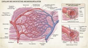
7.3 Blood vessels and cardiovascular system
The cardiovascular system contains several important cell types that can undergo senescence.
These include:
- Endothelial cells lining blood vessels.
- Vascular smooth-muscle cells.
- Fibroblasts surrounding blood vessels.
Their functions include regulating blood flow, maintaining vessel structure and supporting vascular repair.
Senescence-related changes may contribute to vascular inflammation, impaired endothelial function and age-related cardiovascular dysfunction.
7.4 Lungs
Lung tissue contains epithelial cells, fibroblasts, endothelial cells and immune cells.
Senescence in these cells may influence:
- Tissue repair.
- Inflammatory signalling.
- Extracellular matrix remodelling.
- Lung tissue scarring, known as fibrosis.
7.5 Liver
The liver has remarkable regenerative capacity.
Several liver cell populations can enter senescence in response to injury or stress.
In some circumstances, temporary senescence helps control abnormal cellular growth and coordinates repair.
Persistent senescence may contribute to chronic inflammation and fibrosis.
7.6 Kidneys
Kidney cells perform filtration, fluid regulation and biochemical balance.
Senescence in kidney cells can affect tissue maintenance and repair.
Persistent senescence is being investigated in relation to age-associated kidney dysfunction and chronic kidney disease.
7.7 Muscles
Muscle tissue depends on coordinated activity between muscle fibres, satellite cells, fibroblasts, blood vessels and immune cells.
Senescence-related changes in supporting cell populations may influence muscle repair and regeneration.
Importantly, not every non-dividing muscle cell is senescent. Mature muscle fibres are normally specialised cells that do not routinely divide.
Chapter 8. The surprising benefits of zombie cells
One of the biggest misunderstandings about zombie cells is the belief that they are entirely harmful.
That is scientifically incorrect.
Cellular senescence is an important protective mechanism in human biology.
Its effects depend on the circumstances, location and duration.
Frontiers | Senescence in Wound Repair: Emerging Strategies to Target Chronic Healing Wounds
8.1 Protection against cancer
Imagine a cell developing serious genetic damage.
If that cell continues dividing uncontrollably, it could contribute to tumour formation.
Senescence can stop the damaged cell from multiplying.
This is an important tumour-suppressing mechanism.
8.2 Wound healing
During wound repair, temporary senescent cells can release signalling molecules that coordinate:
- Immune-cell recruitment.
- Tissue remodelling.
- Fibroblast activity.
- Regenerative processes.
After their temporary role has been completed, immune mechanisms can help remove them.
8.3 Embryonic development
Cellular senescence also participates in particular developmental processes.
During embryonic development, some cells temporarily enter senescence-like states that help regulate tissue formation and remodelling.
This demonstrates that senescence is not simply a consequence of ageing.
8.4 Tissue organisation
Senescent cells can influence the extracellular matrix, which is the structural material surrounding cells.
This matrix provides:
- Mechanical support.
- Structural organisation.
- Biochemical signals.
- A framework for tissue repair.
8.5 Controlling abnormal cell growth
Senescence can act as a biological brake.
It prevents certain abnormal cells from continuing to multiply.
This is especially important when cellular damage or abnormal growth signals threaten normal tissue organisation.
Chapter 9. The harmful consequences of accumulated zombie cells
The problem arises when senescent cells persist and their secretory activities become chronically disruptive.
9.1 Chronic inflammation
Persistent SASP activity can contribute to prolonged inflammatory signalling.
Inflammation is normally useful in defence and repair.
However, when it continues unnecessarily, it may interfere with normal tissue function.
9.2 Fibrosis
Fibrosis occurs when excessive connective tissue, particularly collagen, accumulates within an organ.
Senescent-cell signalling can influence fibroblast activity and extracellular matrix production.
This may contribute to tissue stiffness and impaired organ function.
9.3 Reduced tissue regeneration
Many tissues depend on stem cells and progenitor cells for maintenance and repair.
A persistently inflammatory environment can interfere with their function.
This may reduce the capacity of tissues to regenerate efficiently.
9.4 Cardiovascular ageing
Senescence in vascular cells can influence blood-vessel function.
Researchers are investigating its relationship with:
- Reduced endothelial function.
- Vascular inflammation.
- Arterial stiffness.
- Atherosclerosis-related processes.
9.5 Cancer-related effects
Senescence has a complex relationship with cancer.
Initially, it can prevent cancer by stopping damaged cells from dividing.
However, senescent cells that persist inside an established tumour environment may release signals that support tumour growth, alter surrounding tissues or influence treatment resistance.
This is why scientists distinguish between the protective effects of initiating senescence and the potentially harmful effects of long-term senescent-cell persistence.DOI+2
Chapter 10. Understanding the differences between normal, dormant, senescent and dead cells
This distinction is fundamental to understanding human cellular biology.
| Characteristic | Normal cell | Quiescent cell | Senescent cell | Dead cell |
|---|---|---|---|---|
| Alive | Yes | Yes | Yes | No |
| Metabolically active | Yes | Yes | Yes | No sustained cellular metabolism |
| Divides | Depending on cell type | Can re-enter division | Usually persistently arrested | No |
| Performs specialised functions | Yes | Yes | Often altered | No |
| Produces signalling molecules | Yes | Yes | Often increased SASP | Not as an active cell |
| Can return to normal growth | Yes | Often yes | Usually not | No |
What is quiescence?
Quiescence is a reversible resting state.
A quiescent cell is not actively dividing, but it can sometimes return to the cell cycle when the body requires it.
For example, certain stem cells remain quiescent until tissue repair is necessary.
What is apoptosis?
Apoptosis is programmed cell death.
It is a controlled biological process through which the body eliminates cells that are no longer required or are damaged.
What makes senescence different?
Senescence is characterised by persistent cell-cycle arrest while the cell remains alive and metabolically active.
This is the defining distinction behind the zombie-cell nickname.
Chapter 11. How scientists identify zombie cells
Scientists cannot reliably identify senescent cells simply by looking at a tissue sample.
They use several complementary laboratory methods.
Checking your browser – reCAPTCHA
| Scientific marker | What researchers investigate |
|---|---|
| p16INK4a | Cell-cycle inhibition |
| p21CIP1 | Cell-cycle arrest |
| SA-β-gal | Increased lysosomal enzyme activity |
| γH2AX | DNA damage responses |
| Lamin B1 | Changes in nuclear structure |
| Ki-67 | Cellular proliferation |
| IL-6 and IL-8 | Inflammatory signalling |
| SASP proteins | Senescence-associated secretion |
Why multiple markers are necessary
There is no single universal marker that conclusively identifies every senescent cell.
For example:
- p21 can increase in cells experiencing temporary stress.
- SA-β-gal activity can occur in certain non-senescent cells.
- DNA damage is not exclusive to senescence.
- Inflammatory molecules can be produced by normal immune cells.
Scientists therefore combine molecular markers, cell-cycle measurements, tissue location and other characteristics.
This is particularly important when studying human organs.PubMed Central (PMC)+1
Chapter 12. The future of zombie-cell research: Senolytics and senomorphics
Modern biotechnology is investigating whether it is possible to reduce the harmful effects of persistent senescent cells.
Two important research areas are senolytics and senomorphics.
T cells engineered to target senescence | Nature
12.1 Senolytics
Senolytics are experimental therapeutic approaches designed to selectively eliminate certain senescent cells.
The underlying scientific idea is to identify biological survival mechanisms that senescent cells depend on and investigate whether these can be targeted.
The objective is not to eliminate every senescent cell.
That would be undesirable because some senescent cells perform beneficial functions.
12.2 Senomorphics
Senomorphics are approaches intended to modify the harmful behaviour of senescent cells without necessarily eliminating them.
Researchers investigate whether it is possible to reduce harmful SASP signalling and associated inflammation.
12.3 Immune-based approaches
Another important research direction is improving the immune system’s ability to recognise and remove persistent senescent cells.
Scientists are investigating:
- Natural killer cell activity.
- Macrophage function.
- T-cell surveillance.
- Senescence-associated surface proteins.
- Immune recognition mechanisms.
12.4 What is the current scientific position?
These are active areas of biomedical research, not established general anti-ageing treatments.
There are important questions about safety, tissue specificity, long-term effects and the preservation of beneficial senescent cells.
People should not attempt to eliminate senescent cells using unproven supplements or self-administered experimental substances.
Chapter 13. The relationship between zombie cells and human DNA
To understand senescent cells properly, we must connect three major biological systems:
- The human genome.
- The cellular machinery.
- The immune system.
【Essential 細胞生物学】1章 細胞とはPart 1|タックスヘイヴン
13.1 DNA: The information-storage system
DNA stores the genetic instructions needed for cellular development, maintenance and reproduction.
Senescence changes how the cell reads and uses certain genetic instructions.
The DNA itself is not necessarily destroyed when senescence develops.
Instead, changes in gene regulation influence which proteins the cell produces.
13.2 RNA: The information-processing system
RNA molecules help transfer and regulate genetic information.
Messenger RNA (mRNA), for example, carries instructions used by ribosomes to manufacture proteins.
Senescent cells change their patterns of RNA production.
This allows them to produce different combinations of proteins, including proteins involved in inflammation, stress responses and cellular survival.
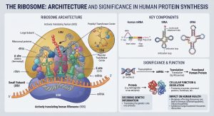
13.3 Ribosomes: The protein-manufacturing system
Ribosomes translate mRNA into proteins.
Senescent cells continue producing proteins, but their protein-production patterns change.
Some proteins support cell survival, while others contribute to SASP signalling.
13.4 Proteins: The operational machinery
Proteins carry out most cellular functions.
In senescent cells, changes in protein expression influence:
- Cell-cycle control.
- DNA repair.
- Inflammatory signalling.
- Mitochondrial activity.
- Cellular communication.
- Resistance to cell death.
The relationship can be summarised as:
DNA
Genetic information
RNA
Genetic information processing
Ribosomes
Protein manufacturing
Proteins
Cellular operations and communication
Senescence-associated changes
Altered metabolism, cell division and signalling
Chapter 14. The scientific architecture of the entire process
The following diagram connects the major mechanisms discussed throughout this thesis.
The biological process can be understood through six stages:
| Stage | Biological event | Significance |
|---|---|---|
| 1 | Cellular stress | DNA damage, telomere dysfunction or oxidative stress |
| 2 | Molecular detection | Damage-response pathways become activated |
| 3 | Growth arrest | p53-p21 and p16-RB pathways restrict division |
| 4 | Senescent state | Cell remains alive but changes its activity |
| 5 | SASP signalling | Cell communicates with surrounding tissues |
| 6 | Final outcome | Immune clearance or persistent senescence |
The final outcome is particularly important.
A temporary senescent state can contribute to healthy tissue repair. Persistent senescence can contribute to inflammation and tissue dysfunction.
Chapter 15. New scientific developments: Mapping zombie cells throughout the human body
An important development was reported by the US National Institutes of Health in 2026.
Researchers associated with the Cellular Senescence Network (SenNet) have been investigating the location, molecular characteristics and diversity of senescent cells throughout the human body.
One development is the Senescence Catalog (SenCat), which includes gene-activity and protein information from 14 types of human senescent cells.
Researchers identified different molecular patterns, demonstrating that senescent cells do not all behave identically.
This is important because it suggests that future research may need to identify particular senescent-cell populations rather than treating all senescent cells as one biological category.
It could help scientists develop more precise approaches to understanding cellular ageing and tissue dysfunction.
Source: NIH – Senescent cells mapped in the human body over the lifespan.
Chapter 16. The economic and technological significance of zombie-cell research
Cellular senescence is not only a subject of biological interest. It is also becoming important to biotechnology, pharmaceutical research and the future healthcare economy.
Several scientific industries are involved.
| Industry | Contribution |
|---|---|
| Genomics | Studies genetic changes associated with senescence |
| Proteomics | Studies proteins produced by senescent cells |
| Biotechnology | Develops tools for investigating cellular behaviour |
| Pharmaceutical research | Investigates potential senescence-related treatments |
| Artificial intelligence | Analyses complex biological datasets |
| Medical imaging | Investigates tissue and cellular changes |
| Computational biology | Models molecular interactions and biological pathways |
The role of artificial intelligence
AI can help researchers investigate senescence by:
- Analysing large genomic datasets.
- Identifying gene-expression patterns.
- Comparing senescent cells from different organs.
- Analysing protein interactions.
- Modelling cellular communication.
- Identifying potential biological targets for further research.
However, computational predictions must be tested through biological experiments and clinical research.
This is an excellent example of how mathematics, physics, computer science, genetics and biochemistry are becoming interconnected in modern medical innovation.
Chapter 17. Important scientific questions for future research
The following questions represent major research challenges:
- Why do certain cells enter senescence earlier than others?
- Why do some senescent cells escape immune-system clearance?
- How do mitochondria influence the development of senescence?
- How does SASP influence neighbouring healthy cells?
- Can researchers distinguish beneficial senescence from harmful persistent senescence?
- How do senescent cells influence stem-cell regeneration?
- Can artificial intelligence identify senescent cells more accurately?
- Can treatments target harmful senescent cells without interfering with beneficial cellular processes?
- How do senescent cells influence different organs simultaneously?
- What determines whether senescence protects against cancer or contributes to an established tumour environment?
These questions demonstrate why cellular senescence is an important field of modern molecular medicine.
Chapter 18. Final scientific conclusion
The so-called zombie cell is one of the most interesting discoveries in modern cellular biology because it challenges our traditional understanding of what it means for a cell to be alive, functional and healthy.
A senescent cell is neither an ordinary dividing cell nor a dead cell.
It is a living biological structure that has entered a persistent state of growth arrest while retaining metabolic activity and the ability to communicate with its surroundings.
Its architecture involves the coordinated activity of:
- DNA and chromosomes.
- Nuclear regulatory proteins.
- p53, p21, p16 and RB.
- Mitochondria and cellular metabolism.
- Ribosomes and protein production.
- Lysosomes and cellular recycling.
- SASP signalling molecules.
- Immune-system surveillance.
The most important discovery is that senescence has two contrasting biological consequences.
First: Protection.
It prevents certain damaged cells from multiplying, supports aspects of wound healing and participates in embryonic development.
Second: Potential damage.
When senescent cells persist, their inflammatory secretions and altered metabolic activities can contribute to tissue ageing, fibrosis and age-associated disease.
The future of this science therefore does not simply involve eliminating zombie cells. It involves understanding their molecular identities, biological functions, communication systems and appropriate removal by the immune system.
The ultimate scientific objective is to understand how the human body maintains a healthy balance between cellular protection, regeneration, repair and the removal of damaged cells.
Recommended scientific reading and research references
These publications provide a foundation for further study.
1. Nature Reviews Molecular Cell Biology (2024). The Senescence-Associated Secretory Phenotype and Its Physiological and Pathological Implications. Nature
2. Experimental & Molecular Medicine (2025). Hallmarks of Cellular Senescence: Biology, Mechanisms, Regulations. Nature
3. SenNet Recommendations for Detecting Senescent Cells in Different Tissues. PubMed Central (PMC)
4. Nature Aging (2025). Immune Surveillance of Senescent Cells in Aging and Disease. Nature
5. National Institutes of Health (2026). Senescent Cells Mapped in the Human Body Over the Lifespan. National Institutes of Health (NIH)




Be First to Comment