
COMPREHENSIVE SCIENTIFIC THESIS
Abstract
The intestinal villi are among the most important microscopic structures in the human digestive system. They are specialised, finger-like projections extending from the inner lining of the small intestine. Their primary responsibility is to absorb nutrients produced during digestion and transfer those nutrients into the body’s circulation.
Without intestinal villi, the human body would struggle to obtain the essential nutrients required for energy production, cellular construction, tissue repair, immune function, brain activity, and general survival.
However, villi are much more than simple absorption structures. They form an extraordinarily organised biological interface involving epithelial cells, blood capillaries, lymphatic vessels, immune cells, enzymes, transport proteins, and specialised cellular junctions.
This thesis investigates their complete microscopic architecture, structural characteristics, physiological mechanisms, relationship with other digestive organs, and importance to the modern understanding of human biology.
Chapter 1. Understanding the small intestine
The small intestine is a long, specialised digestive organ situated between the stomach and the large intestine.
Its major responsibilities include:
- Completing the chemical digestion of food.
- Absorbing carbohydrates.
- Absorbing proteins and amino acids.
- Absorbing fats and fatty acids.
- Absorbing vitamins and minerals.
- Absorbing water and electrolytes.
- Supporting the immune system.
- Communicating with the nervous and hormonal systems.
The small intestine is divided into three anatomical regions.
| Region | Principal function |
|---|---|
| Duodenum | Receives stomach contents, bile and pancreatic secretions; continues digestion |
| Jejunum | Performs a substantial proportion of nutrient absorption |
| Ileum | Absorbs remaining nutrients, including vitamin B12 and recycled bile salts |
The intestinal villi occur throughout the small intestine, although their characteristics and distribution vary between regions.
An important scientific principle: The small intestine does not depend on one structure alone. Its digestive efficiency comes from the coordinated operation of folds, villi, microvilli, digestive enzymes, transport proteins, blood vessels, and lymphatic vessels.
Chapter 2. What exactly are intestinal villi?
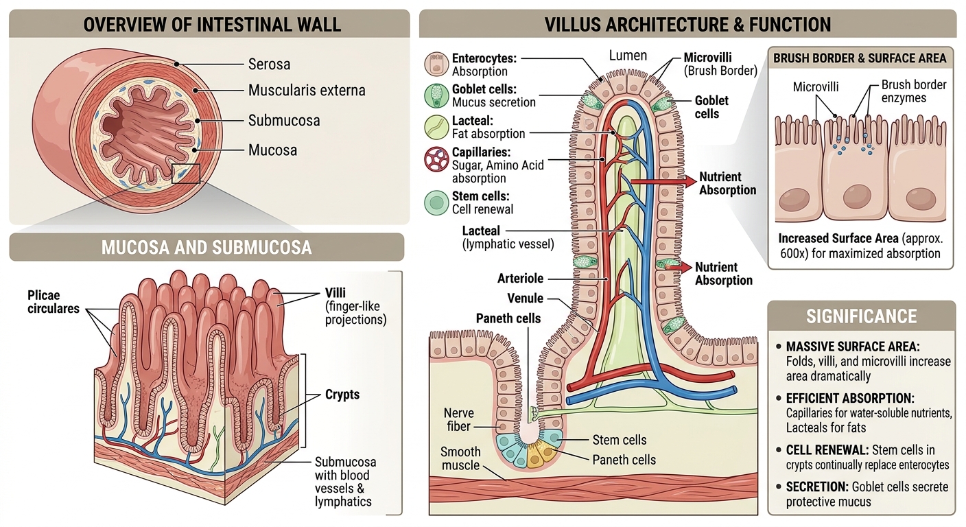
A villus (plural: villi) is a microscopic, finger-shaped projection of the intestinal mucous membrane.
Each villus projects into the intestinal lumen, the internal space through which partially digested food travels.
Its architecture increases the available surface area for nutrient absorption.
Imagine a flat surface compared with a surface covered in thousands of tiny projections. The projected surface offers considerably more area through which materials can be absorbed.
This is the fundamental engineering principle behind intestinal villi.
The three levels of surface-area organisation
The small intestine uses three interconnected structural levels:
| Structure | Anatomical description | Main contribution |
|---|---|---|
| Circular folds | Large internal folds of the intestinal wall | Increase surface area and slow movement of intestinal contents |
| Villi | Finger-like projections of the mucosa | Provide additional absorptive surface |
| Microvilli | Microscopic projections on individual epithelial cells | Create the brush border and increase absorption capacity |
Microvilli are considerably smaller than villi. They are found on the surface of the absorptive epithelial cells called enterocytes.
Together, these structures create a highly efficient biological absorption system.
Chapter 3. Complete microscopic architecture of an intestinal villus
This is the central chapter for understanding how villi operate.
A typical intestinal villus contains several interconnected anatomical components.
3.1 The epithelial covering
The outside of each villus is covered by a single layer of specialised epithelial cells.
These cells form the interface between the intestinal contents and the internal environment of the human body.
The epithelium contains several important cell populations.
A. Enterocytes
Enterocytes are the principal absorptive cells.
Their responsibilities include:
- Absorbing glucose and other simple sugars.
- Absorbing amino acids and small peptides.
- Absorbing fatty-acid digestion products.
- Transporting vitamins and minerals.
- Producing digestive enzymes located on their brush border.
- Controlling the movement of selected substances across the intestinal barrier.
Each enterocyte possesses microvilli on its exposed surface.
B. Goblet cells
Goblet cells produce mucus.
Mucus forms a protective and lubricating layer that helps protect the intestinal lining and allows intestinal contents to move more smoothly.
C. Enteroendocrine cells
These specialised cells release hormones in response to nutrients and other intestinal signals.
Examples include:
- Cholecystokinin (CCK).
- Secretin.
- Glucose-dependent insulinotropic polypeptide (GIP).
- Glucagon-like peptide-1 (GLP-1).
These hormones help coordinate digestion, pancreatic secretion, gallbladder activity, and metabolic responses.
D. Tuft cells
Tuft cells are specialised sensory epithelial cells involved in detecting environmental signals and supporting immune responses.
E. Antimicrobial secretory cells
Paneth cells are particularly concentrated in the intestinal crypts at the bases of the villi. They release antimicrobial substances that help regulate intestinal microorganisms.
3.2 The lamina propria
Beneath the epithelial covering is a specialised connective-tissue region called the lamina propria.
It forms the internal supporting framework of the villus.
It contains:
- Blood capillaries.
- Lymphatic vessels.
- Immune cells.
- Fibroblasts.
- Connective-tissue fibres.
- Small nerve fibres.
- Smooth-muscle fibres.
The lamina propria provides structural support and connects absorption with circulation and immune defence.
3.3 Blood capillary network
Each villus contains an extensive network of tiny blood vessels called capillaries.
These vessels receive water-soluble nutrients absorbed through the epithelial cells.
Important transported substances include:
- Glucose.
- Galactose.
- Amino acids.
- Water-soluble vitamins.
- Many minerals.
The absorbed nutrients enter the bloodstream and travel through the hepatic portal circulation towards the liver.
The liver subsequently processes, stores, transforms, and distributes many of these nutrients.
3.4 The central lacteal
One of the most interesting structures inside an intestinal villus is the lacteal.
A lacteal is a specialised lymphatic capillary located near the centre of the villus.
Its primary function is absorbing dietary fats packaged into particles called chylomicrons.
The process works as follows:
- Dietary fats are digested into smaller components.
- These components enter intestinal epithelial cells.
- Many long-chain fatty acids are reassembled into triglycerides.
- The triglycerides are packaged with proteins and other lipids into chylomicrons.
- Chylomicrons enter the lacteals.
- They travel through the lymphatic circulation.
- They eventually enter the bloodstream.
This means that the villus has two important transport networks: a blood circulation pathway and a lymphatic pathway.
3.5 Smooth-muscle fibres
Small extensions of smooth muscle from the muscularis mucosae enter the villi.
They help produce local movements of the villus, supporting contact between intestinal contents and the absorptive surface.
Chapter 4. Microvilli: the microscopic machinery of absorption
Microvilli represent an even smaller level of biological engineering.
They are tiny extensions of the plasma membrane covering the upper surface of enterocytes.
Their architecture contains:
- A plasma membrane surrounding each projection.
- Bundles of actin filaments forming the internal structural framework.
- Membrane proteins responsible for nutrient transport.
- Digestive enzymes attached to the brush-border membrane.
- Glycoproteins that contribute to the protective glycocalyx.
4.1 The brush-border system
Thousands of microvilli are organised together to form what scientists call the brush border.
This is where the final stages of carbohydrate and protein digestion occur.
Examples of brush-border enzymes include:
| Enzyme | Function |
|---|---|
| Maltase | Breaks maltose into glucose |
| Sucrase | Breaks sucrose into glucose and fructose |
| Lactase | Breaks lactose into glucose and galactose |
| Peptidases | Break small peptides into amino acids and smaller peptides |
| Isomaltase | Helps digest branched carbohydrate products |
The brush border therefore performs two closely connected operations:
- Final chemical digestion.
- Absorption of the resulting nutrients.
This arrangement is highly efficient because digestion and absorption occur in close proximity.
Chapter 5. The complete nutrient absorption mechanism
The most important responsibility of intestinal villi is transferring nutrients from digested food into the internal transport systems of the human body.
Let us examine how this works at the molecular level.
5.1 Carbohydrate absorption
Carbohydrates provide an important source of energy.
However, large carbohydrate molecules cannot simply pass directly into the bloodstream.
They must first be broken down into smaller molecules called monosaccharides.
The three principal monosaccharides are:
- Glucose.
- Galactose.
- Fructose.
The absorption mechanism involves specialised membrane transport proteins.
Glucose and galactose
They enter enterocytes mainly through a transporter called SGLT1.
SGLT1 uses the sodium gradient to transport these sugars into the cell.
The sodium gradient is maintained by the sodium-potassium ATPase pump on the opposite side of the enterocyte.
Fructose
Fructose enters through a different transporter called GLUT5.
Exit into circulation
The absorbed monosaccharides leave the enterocyte mainly through GLUT2 on its blood-facing membrane.
They then enter the villus capillaries and travel towards the liver.
This is an excellent example of how microscopic biological structures use specialised molecular transport machinery.
5.2 Protein and amino-acid absorption
Proteins are essential for constructing and maintaining human tissues.
Digestive enzymes break dietary proteins into:
- Amino acids.
- Dipeptides.
- Tripeptides.
These products are absorbed by enterocytes through different transport systems.
A particularly important transporter is PEPT1, which absorbs many dipeptides and tripeptides using a hydrogen-ion gradient.
Inside the enterocyte, most absorbed small peptides are broken down into individual amino acids.
The amino acids then leave the cells and enter the blood capillaries.
The liver and other tissues use these amino acids to manufacture proteins required for:
- Muscle and tissue maintenance.
- Enzymes.
- Hormones.
- Antibodies.
- Structural proteins.
- Cellular repair.
5.3 Fat absorption
Fat absorption uses a different transportation architecture from carbohydrate and protein absorption.
The process involves several stages.
Stage 1: Emulsification
Bile salts produced by the liver and released through the biliary system help disperse large fat droplets into smaller droplets.
Stage 2: Enzymatic digestion
Pancreatic lipase breaks triglycerides into smaller lipid products, principally free fatty acids and monoglycerides.
Stage 3: Micelle formation
Bile salts help form micelles that transport lipid digestion products towards the epithelial surface.
Stage 4: Entry into enterocytes
Lipid components enter the intestinal epithelial cells.
Stage 5: Reassembly
Inside the enterocyte, many long-chain fatty acids and monoglycerides are reassembled into triglycerides.
Stage 6: Chylomicron formation
The triglycerides are packaged with cholesterol, phospholipids, and specialised proteins into chylomicrons.
Stage 7: Entry into the lacteal
Chylomicrons enter the lymphatic capillaries inside the villi.
Stage 8: Distribution
The lymphatic system transports them towards the bloodstream, where their lipid contents can be distributed to tissues.
An important exception is that many short- and medium-chain fatty acids can enter the portal blood directly rather than following the usual chylomicron-lacteal pathway.
5.4 Vitamin absorption
Vitamins are essential micronutrients involved in hundreds of biological processes.
The intestinal villi provide the absorption interface for these nutrients.
| Vitamin | Principal biological importance |
|---|---|
| A | Vision, epithelial maintenance and immune function |
| B1 | Energy metabolism and nerve function |
| B2 | Cellular energy metabolism |
| B3 | Metabolic reactions |
| B6 | Amino-acid metabolism and nervous-system function |
| B9 | DNA synthesis and cell division |
| B12 | Red blood cell formation and nervous-system function |
| C | Collagen synthesis and antioxidant functions |
| D | Calcium and phosphate regulation |
| E | Protection of cell membranes from oxidative damage |
| K | Normal blood clotting and bone-related processes |
Fat-soluble vitamins A, D, E and K generally travel with dietary lipids and are incorporated into chylomicrons.
Water-soluble vitamins use specific transport proteins or other specialised absorption mechanisms.
Vitamin B12 is particularly interesting because its absorption requires intrinsic factor, a protein produced by the stomach, and specialised receptors in the terminal ileum.
5.5 Mineral absorption
The small intestine also absorbs essential minerals.
Examples include:
- Iron.
- Calcium.
- Magnesium.
- Zinc.
- Phosphorus.
- Copper.
- Selenium.
Different minerals have different absorption mechanisms.
For example, iron absorption occurs primarily in the duodenum and upper jejunum, while calcium absorption is regulated partly by vitamin D.
Chapter 6. The two transportation systems inside the villus
The villus is particularly remarkable because it contains two distinct transport networks.
6.1 The blood circulation network
The blood capillaries collect most water-soluble nutrients.
Their general pathway is:
Intestinal lumen
Digested nutrients
Enterocytes
Nutrient absorption
Villus blood capillaries
Hepatic portal vein
Liver
Processing, storage and metabolic regulation
Systemic circulation
Distribution throughout the body
6.2 The lymphatic transportation network
The lymphatic network primarily receives absorbed long-chain dietary fats packaged into chylomicrons.
Intestinal enterocytes
Chylomicron production
Central lacteal
Intestinal lymphatic vessels
Thoracic duct
Venous circulation
Entry into the bloodstream
These two systems work together to distribute the products of digestion.
Chapter 7. Intestinal villi and the human immune system
The intestinal lining is not merely a nutrient absorption surface.
It is also an important immunological defence boundary.
The intestine encounters enormous numbers of microorganisms, food-derived substances and environmental molecules.
Its immune system must distinguish between:
- Nutrients that should be absorbed.
- Beneficial microorganisms that can coexist with the body.
- Potentially harmful microorganisms.
- Substances that may trigger inappropriate immune responses.
Important immune components
1. Intraepithelial lymphocytes
These immune cells are located between intestinal epithelial cells and contribute to local immune surveillance.
2. T lymphocytes
They coordinate immune responses and help maintain immune balance.
3. B lymphocytes and plasma cells
Plasma cells produce antibodies, including immunoglobulin A (IgA), which helps protect mucosal surfaces.
4. Macrophages
They help remove unwanted microorganisms and cellular debris.
5. Dendritic cells
They collect and present antigens to other immune cells, helping regulate immune responses.
6. Peyer’s patches
These are organised immune structures particularly prominent in the ileum. They contain specialised M cells that transport selected material from the intestinal lumen to underlying immune tissue.
An important distinction is that Peyer’s patches are not villi. They are specialised lymphoid structures associated with the intestinal mucosa.
The significance of this entire system is that the intestine must absorb nourishment while maintaining protection against infection.
Chapter 8. The biological renewal and repair architecture
One of the most impressive characteristics of intestinal tissue is its ability to continually renew itself.
The intestinal epithelial lining experiences constant exposure to:
- Digestive enzymes.
- Mechanical movement.
- Food particles.
- Microorganisms.
- Chemical substances.
- Environmental changes.
Consequently, epithelial cells require continuous replacement.
8.1 The intestinal crypts
At the base of each villus are small tubular depressions called intestinal crypts or crypts of Lieberkühn.
These structures contain specialised stem cells.
The stem cells divide and produce daughter cells that develop into different intestinal epithelial cell types.
8.2 Cell differentiation
New cells develop into specialised populations, including:
- Enterocytes.
- Goblet cells.
- Enteroendocrine cells.
- Paneth cells.
Enterocytes and several other epithelial cell populations migrate towards the villus tip.
Paneth cells are an important exception: they generally migrate towards and remain near the bottom of the crypts.
8.3 Cellular replacement
Mature intestinal epithelial cells are eventually shed and replaced.
This continuous renewal helps maintain the intestinal barrier and its absorptive functions.
The intestinal lining therefore represents a dynamic biological structure rather than a permanently fixed surface.
The crypt-villus arrangement is an excellent example of stem-cell biology, cellular differentiation, tissue organisation and biological regeneration operating together.
Chapter 9. The nervous and hormonal control of intestinal villi
The villi do not operate independently.
Their activities are coordinated by the nervous system, digestive hormones, local chemical signals and the circulation.
9.1 The enteric nervous system
The enteric nervous system (ENS) is a network of neurons embedded within the digestive tract.
It is sometimes called the gut’s intrinsic nervous system.
It regulates:
- Intestinal movement.
- Local blood flow.
- Digestive secretions.
- Coordination of absorption and digestion.
- Communication between different intestinal regions.
Two important nerve networks are:
| Network | Location | Function |
|---|---|---|
| Myenteric plexus | Between circular and longitudinal muscle layers | Regulates intestinal movement |
| Submucosal plexus | Within the submucosa | Regulates secretion, local blood flow and mucosal activity |
9.2 Hormonal regulation
The small intestine contains specialised endocrine cells that release hormones.
| Hormone | Main responsibility |
|---|---|
| Secretin | Stimulates bicarbonate secretion from the pancreas |
| CCK | Stimulates pancreatic enzyme secretion and gallbladder contraction |
| GIP | Participates in glucose-dependent insulin release |
| GLP-1 | Supports glucose regulation and influences gastric emptying |
| Motilin | Helps regulate migrating motor activity between meals |
| PYY | Contributes to appetite regulation and gastrointestinal motility |
The importance of these hormones is that digestion, nutrient absorption and metabolism are coordinated rather than operating as isolated processes.
Chapter 10. The relationship between intestinal villi and cellular energy
This is an especially important connection between intestinal anatomy and biochemistry.
The villi absorb nutrients, but the body’s cells must convert those nutrients into usable biological energy.
The principal energy-carrying molecule in cells is ATP (adenosine triphosphate).
How the relationship works
- Villi absorb glucose, amino acids, fatty acids and other nutrients.
- Blood and lymphatic circulation transport these substances.
- Nutrients reach different organs and tissues.
- Cells use metabolic pathways to process these substances.
- Mitochondria generate substantial amounts of ATP through oxidative phosphorylation.
ATP supports:
- Muscle contraction.
- Nerve signalling.
- Active transport across cell membranes.
- Protein synthesis.
- Cellular maintenance.
- Tissue repair.
- Chemical reactions necessary for life.
There is an additional connection: intestinal epithelial cells themselves also require ATP.
For example, the sodium-potassium ATPase pump consumes ATP to maintain the sodium gradient used by SGLT1 during glucose and galactose absorption.
This illustrates a fundamental biological principle:
The intestinal villi absorb the raw materials, while cellular metabolic systems convert and utilise those materials to support life.
Chapter 11. The significance of villi in human survival
The importance of intestinal villi becomes clearer when we examine the major systems that depend on nutrient absorption.
11.1 Brain and nervous system
Absorbed nutrients support brain metabolism, neurotransmitter production, nerve-cell maintenance and normal nervous-system activity.
11.2 Muscular system
Amino acids support muscle protein maintenance, while glucose and fatty acids contribute to energy production.
11.3 Skeletal system
Calcium, phosphorus, vitamin D and protein support bone development and maintenance.
11.4 Cardiovascular system
Absorption of iron, vitamin B12, folate and other nutrients supports normal blood-cell production and cardiovascular function.
11.5 Immune system
Nutrients support immune-cell production, antibody synthesis and normal immune responses.
11.6 Skin and connective tissue
Amino acids, vitamin C, zinc and other nutrients contribute to collagen production, skin maintenance and tissue repair.
11.7 Endocrine and metabolic systems
Nutrient absorption supplies materials for metabolic regulation, hormone synthesis and normal cellular functions.
The villi are therefore connected indirectly to nearly every major organ system.
Chapter 12. What happens when intestinal villi become damaged?
Healthy villi are essential for maintaining normal intestinal absorption.
When their structure becomes damaged, the intestine may lose part of its absorptive capacity.
One recognised example is coeliac disease.
This is an autoimmune condition in which exposure to gluten in genetically susceptible individuals triggers an immune response that damages the small-intestinal lining.
Possible consequences include:
- Villous atrophy.
- Reduced nutrient absorption.
- Iron deficiency.
- Vitamin deficiencies.
- Diarrhoea or digestive discomfort.
- Impaired growth and development in affected children and adolescents.
Other conditions associated with intestinal villous damage or malabsorption include certain intestinal infections and immune-related disorders.
Understanding villous atrophy
Villous atrophy refers to the shortening or flattening of intestinal villi.
The consequences can include:
- Reduced absorptive surface area.
- Disruption of the epithelial barrier.
- Changes in digestive enzyme activity.
- Reduced nutrient transport.
- Nutritional deficiencies.
It is important to understand that not every digestive problem is caused by damaged villi. Some conditions affect pancreatic enzymes, bile production, intestinal movement or nutrient transport without directly destroying the villous architecture.
Chapter 13. The intestinal microbiome and villi
The human intestine contains a complex community of microorganisms known as the gut microbiome.
These include bacteria and other microorganisms.
The microbiome interacts with the intestinal lining through chemical signals and metabolic products.
Some of its important contributions include:
- Fermentation of dietary fibre.
- Production of short-chain fatty acids.
- Interaction with immune cells.
- Support of intestinal barrier functions.
- Modification of the intestinal chemical environment.
Short-chain fatty acids, particularly butyrate, are important energy sources for colonocytes, the epithelial cells lining the large intestine.
The relationship between the microbiome and small-intestinal villi is complex. Microorganisms can influence intestinal immune activity, epithelial development and barrier function.
However, the microbiome does not replace the villi’s own absorptive machinery.
Chapter 14. The complete biological engineering model
Let us now combine the entire architecture into one functional system.
The small intestine can be understood as a coordinated biological processing and distribution network.
| Biological component | Engineering equivalent | Actual responsibility |
|---|---|---|
| Circular folds | Surface expansion | Increase internal surface area |
| Villi | Absorption modules | Increase absorptive surface |
| Microvilli | Microscopic interface arrays | Increase membrane area |
| Enterocytes | Processing units | Digest and absorb nutrients |
| Transport proteins | Molecular transport channels | Move specific substances |
| Tight junctions | Selective barriers | Regulate movement between cells |
| Blood capillaries | Distribution network | Transport water-soluble nutrients |
| Lacteals | Specialised distribution network | Transport dietary fats |
| Crypt stem cells | Regeneration system | Replace intestinal epithelial cells |
| Immune cells | Biological security system | Protect intestinal tissues |
| Enteric neurons | Control network | Coordinate digestive activities |
| Enteroendocrine cells | Chemical signalling system | Coordinate digestive and metabolic responses |
This comparison demonstrates how biological systems achieve complex functions through the coordination of many specialised structures.
Chapter 15. Scientific research: important discoveries about intestinal villi
The study of intestinal villi connects several major scientific disciplines.
| Scientific discipline | What it investigates |
|---|---|
| Anatomy | Physical organisation of intestinal structures |
| Histology | Microscopic tissue architecture |
| Cell biology | Enterocyte structure and cellular renewal |
| Molecular biology | Transport proteins and cellular signalling |
| Biochemistry | Enzymes, digestion and nutrient metabolism |
| Immunology | Intestinal immune defence |
| Neuroscience | Enteric nervous system |
| Microbiology | Gut microorganisms |
| Physiology | Nutrient absorption and intestinal function |
| Genetics | Genetic regulation of intestinal development and disease |
Five particularly important scientific concepts
1. Surface-area amplification: Circular folds, villi and microvilli dramatically increase the available absorptive surface. Some educational anatomical references describe an increase exceeding 600-fold compared with a smooth tube, although estimates vary according to the measurement method.
2. Membrane transport: Nutrient absorption depends on specialised molecular transporters rather than simple unrestricted passage through cell membranes.
3. Stem-cell regeneration: Intestinal crypts contain stem cells that continually produce replacement epithelial cells.
4. Dual circulation: Blood capillaries and lymphatic lacteals provide different routes for distributing absorbed nutrients.
5. Immune integration: The intestinal lining is simultaneously an absorptive surface and an important immune barrier.
Chapter 16. Final scientific conclusions
The intestinal villus is an extraordinary example of biological organisation.
Its architecture demonstrates how microscopic structures can perform sophisticated functions through coordinated cellular activities.
Consider what happens when you eat a normal meal:
- The stomach begins mechanical and chemical digestion.
- The pancreas supplies digestive enzymes.
- The liver produces bile.
- The small intestine completes much of the digestion.
- The villi absorb the resulting nutrients.
- The enterocytes transport nutrients across their membranes.
- Blood capillaries carry many nutrients towards the liver.
- Lacteals transport much of the absorbed dietary fat.
- The circulation distributes nutrients throughout the body.
- Individual cells use these nutrients for energy, growth, maintenance and repair.
The entire process is interconnected.
The central biological principle
The villi are the microscopic gateway between the external nutritional environment and the internal biological systems of the human body.
Their importance is not simply that they absorb food. They convert the products of digestion into a form that can enter the body’s transport systems and support the functioning of almost every organ.
Recommended scientific reference library
For further academic study, these are useful research and educational references:
- NCBI Bookshelf — Physiology of the Small Bowel.
- NCBI Bookshelf — Physiology of Nutrient Absorption.
- Exploring the Villus — microscopic structure and function.
- OpenStax: The Small and Large Intestines.
- Physiology of Intestinal Absorption and Secretion.
Suggested next scientific research topic
A particularly valuable continuation would be a comprehensive study of the human intestinal epithelial cell (enterocyte), examining its complete molecular architecture, mitochondria, cell membrane, transport proteins, enzymes, DNA, ATP production and relationship with the villi.
Understanding the enterocyte would take your investigation from the anatomical level of the villus down to the molecular machinery operating inside individual cells.

Be First to Comment