
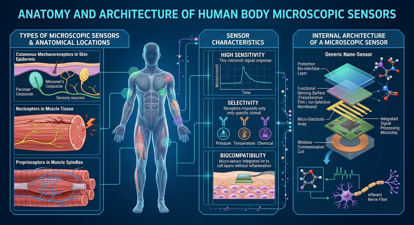
The Anatomy of Microscopic Sensors in the Human Body: Their Architecture, Characteristics, Functions and Significance in Human Survival
HUMAN BIOLOGY • NEUROSCIENCE • CELLULAR BIOLOGY • BIOPHYSICS • PHYSIOLOGY
9.2: Somatosensory Receptors – Social Sci LibreTexts
Dorsal Root Ganglion: Your #1 Pain Breakthrough
ThermoTRP channels and beyond: mechanisms of temperature sensation | Nature Reviews Neuroscience
10
Abstract
The human body contains an extraordinarily sophisticated biological sensing network. Across the skin, muscles, joints, eyes, ears, nose, tongue, blood vessels and internal organs, specialised microscopic structures continuously detect changes in the surrounding environment and within the body itself.
These structures are called sensory receptors.
They function similarly to microscopic biological sensors in an advanced technological system. They detect physical, chemical and biological changes and convert those changes into electrical or chemical signals that the nervous system can interpret.
For example:
- Skin receptors detect touch, pressure, vibration and temperature.
- Muscle receptors detect stretching and changes in muscle length.
- Joint receptors provide information about body position.
- Eye receptors detect light.
- Ear receptors detect sound vibrations and head movement.
- Chemical receptors monitor oxygen, carbon dioxide and other substances.
- Internal receptors detect changes in blood pressure, organ expansion and internal chemical conditions.
The remarkable feature is that these sensors operate as an integrated network rather than as isolated structures.
This thesis investigates their microscopic anatomy, molecular architecture, operating mechanisms, characteristics, communication pathways and contribution to human survival.
CHAPTER 1: Understanding the concept of biological microscopic sensors
1.1 What are microscopic sensors?
A microscopic biological sensor is a specialised cell, nerve ending or cellular structure that detects a particular stimulus and initiates a biological response.
The scientific term is sensory receptor.
A stimulus is any detectable change that can activate a receptor.
Examples include:
| Stimulus | Biological sensor | Information detected |
|---|---|---|
| Pressure | Mechanoreceptor | Physical force |
| Temperature | Thermoreceptor | Heat and cold |
| Light | Photoreceptor | Light intensity and wavelength |
| Chemical molecules | Chemoreceptor | Chemical composition |
| Tissue-threatening stimuli | Nociceptor | Potential or actual tissue damage |
| Muscle stretching | Proprioceptor | Muscle length and movement |
| Blood oxygen changes | Chemoreceptor | Respiratory chemical conditions |
These sensors are distributed throughout the body.
1.2 Biological sensors compared with electronic sensors
An electronic sensor and a biological receptor perform similar fundamental functions, although their physical mechanisms are different.
Researchers create an artificial tactile skin that mimics human tactile recognition processes
| Electronic technology | Human biological equivalent |
|---|---|
| Pressure sensor | Mechanoreceptor |
| Temperature sensor | Thermoreceptor |
| Optical sensor | Photoreceptor |
| Chemical sensor | Chemoreceptor |
| Motion sensor | Vestibular receptor |
| Position sensor | Proprioceptor |
| Signal-processing circuit | Neural networks |
| Central processor | Brain and spinal cord |
| Feedback controller | Nervous and endocrine systems |
The human body is not literally a computer. However, this comparison helps explain how biological detection, communication, processing and feedback work together.
CHAPTER 2: The complete architecture of the human sensory network
The human sensory system has several interconnected architectural levels.
Advanced Anatomy & Physiology: Sensory System | ditki medical & biological sciences
2.1 The six architectural levels
Level 1: Stimulus detection
The body encounters a physical or chemical change.
Examples:
- Touching a surface.
- Experiencing environmental temperature changes.
- Detecting light.
- Experiencing changes in blood pressure.
Level 2: Receptor activation
A specialised receptor detects the stimulus.
Level 3: Signal transduction
The receptor converts the stimulus into a biological signal.
This may involve:
- Ion channels opening or closing.
- Changes in electrical voltage.
- Release of chemical messengers.
- Activation of intracellular signalling pathways.
Level 4: Neural transmission
Sensory neurons carry information towards the central nervous system.
Level 5: Information processing
The spinal cord and brain process and integrate incoming signals.
Level 6: Biological response
The nervous system can initiate:
- Muscle movement.
- Changes in posture.
- Withdrawal reflexes.
- Changes in heart rate.
- Sweating.
- Changes in breathing.
This creates an interconnected biological feedback system.
2.2 Overall communication architecture
EXTERNAL OR INTERNAL STIMULUS
Pressure • Temperature • Light • Chemicals • Movement
MICROSCOPIC SENSORY RECEPTORS
Detection and biological transduction
PERIPHERAL SENSORY NEURONS
Electrical signal transmission
SPINAL CORD AND BRAIN
Integration, interpretation and coordination
BIOLOGICAL RESPONSE
Movement • Regulation • Protection • Adaptation
This architecture allows the body to respond to thousands of environmental and internal changes.
CHAPTER 3: The seven major categories of microscopic sensory receptors
The following categories provide a useful scientific framework for understanding the body’s biological sensors.
3.1 Mechanoreceptors – sensors of physical force
Specialized Skin Structures | Nerve Endings and Glands – Integrated Human Anatomy and Physiology
Mechanoreceptors detect mechanical changes.
These include:
- Touch.
- Pressure.
- Vibration.
- Stretching.
- Movement.
- Sound waves.
They are found in the skin, muscles, joints, blood vessels and internal organs.
Four important skin mechanoreceptors
A. Merkel cell-neurite complexes
Location: Basal layer of the epidermis.
Functions:
- Detect sustained light pressure.
- Identify edges and surface shapes.
- Support fine tactile discrimination.
- Help identify textures.
Merkel cells work together with sensory nerve endings to provide detailed touch information.
B. Meissner corpuscles
medihistory.elearning.aum.iml.unibe.ch
Location: Dermal papillae, particularly in fingertips, palms and soles.
Functions:
- Detect light touch.
- Detect movement across the skin.
- Help identify when an object begins slipping.
- Respond to low-frequency vibration.
They are rapidly adapting receptors.
C. Pacinian corpuscles
BioA_3.6.1.2 Receptors – Pacinian Corpuscle | Teaching Resources
Location: Deep dermis and subcutaneous tissue.
Architecture:
The Pacinian corpuscle has a distinctive layered structure resembling an onion.
Its components include:
- A central sensory nerve ending.
- Multiple concentric layers of supporting cells.
- A surrounding connective-tissue capsule.
- A mechanical transmission structure.
Functions:
- Detect deep pressure changes.
- Detect vibration.
- Respond to rapid mechanical changes.
Its layered architecture helps determine how mechanical forces reach the sensory nerve ending.
D. Ruffini endings
Location: Deep dermis and connective tissues.
Functions:
- Detect skin stretching.
- Detect sustained mechanical deformation.
- Contribute to information about finger position.
- Support awareness of hand movement.
These four receptor systems work together to give the brain detailed information about physical contact.NCBI+2
3.2 Thermoreceptors – sensors of temperature
PPT – What is Pain? PowerPoint Presentation, free download – ID:4260038
Thermoreceptors detect changes in temperature.
They are particularly important for temperature regulation and environmental awareness.
There are two principal functional categories:
| Receptor | Function |
|---|---|
| Cold-sensitive receptors | Detect cooling |
| Warm-sensitive receptors | Detect warming |
Many temperature-sensitive nerve endings contain specialised molecular ion channels called transient receptor potential (TRP) channels.
Examples include:
- TRPV1: responds to noxious heat and certain chemical stimuli.
- TRPM8: responds to cooling and menthol.
- TRPA1: participates in detecting certain irritating chemicals and environmental stimuli.
These channels allow physical or chemical changes to influence electrical activity in sensory neurons.
The hypothalamus receives temperature information and coordinates important temperature-regulating responses.
These include sweating, changes in skin blood flow and shivering.
3.3 Nociceptors – sensors of potentially harmful stimuli
Nociceptors detect stimuli that threaten or damage body tissues.
They are commonly found as specialised free nerve endings.
They respond to:
- Excessive heat or cold.
- Strong mechanical pressure.
- Tissue inflammation.
- Certain chemical substances associated with tissue damage.
Two important sensory fibre types are:
| Fibre | Characteristics |
|---|---|
| A-delta | Thinly myelinated and relatively fast |
| C-fibres | Unmyelinated and relatively slow |
Nociception is the nervous system’s detection and processing of potentially harmful stimuli.
Pain is the conscious experience associated with this processing and is influenced by brain activity, emotions and context.
Importance: Nociception helps the body recognise potentially damaging conditions and initiate protective responses.
3.4 Chemoreceptors – sensors of chemical composition
Chemoreceptors detect chemical changes.
They are found in several locations, including:
- Carotid bodies.
- Aortic bodies.
- Brainstem-related respiratory control systems.
- Nose.
- Tongue.
- Gastrointestinal system.
Important chemical sensing functions
A. Oxygen monitoring
Peripheral chemoreceptors, especially the carotid bodies, detect reductions in arterial oxygen.
B. Carbon dioxide monitoring
Central respiratory control mechanisms respond strongly to changes in carbon dioxide through their effects on brain extracellular fluid and cerebrospinal fluid pH.
C. Blood acidity monitoring
Chemical sensing mechanisms detect changes associated with blood pH.
These sensors communicate with respiratory control centres in the brainstem.
The brainstem adjusts breathing activity to help maintain appropriate gas exchange.
3.5 Photoreceptors – microscopic sensors of light
Photoreceptors are specialised cells in the retina.
There are two primary types.
| Characteristics | Rods | Cones |
|---|---|---|
| Primary function | Dim-light vision | Colour and detailed vision |
| Sensitivity | Very high | Lower in dim conditions |
| Colour detection | No individual colour discrimination | Different wavelength sensitivities |
| Distribution | Predominantly peripheral retina | Concentrated in central retina |
Architecture of a photoreceptor
A photoreceptor contains:
- Outer segment.
- Light-sensitive membrane structures.
- Photopigments.
- Inner segment.
- Cell nucleus.
- Synaptic terminal.
The photopigment in rods is rhodopsin.
When light interacts with a photopigment, it initiates a biochemical process called phototransduction.
This changes the electrical state of the photoreceptor and influences the activity of connected retinal neurons.
The resulting information eventually travels through the optic nerve to the brain.
3.6 Proprioceptors – sensors of body position and movement
La proprioception : notre sixième sens – Blue Portance
Proprioceptors provide information about the position and movement of the body.
They are especially important for:
- Balance.
- Walking.
- Coordination.
- Posture.
- Controlled muscle movement.
- Awareness of limb position without looking at it.
Three important receptor systems are:
A. Muscle spindles
These specialised sensory structures are located within skeletal muscles.
They detect changes in muscle length and the speed at which muscle length changes.
B. Golgi tendon organs
These are located in tendons near the muscle-tendon junction.
They detect mechanical tension generated through muscle contraction and other loading conditions.
C. Joint sensory receptors
These occur in joint capsules and associated tissues.
They contribute information about joint movement, position and mechanical conditions.
The brain combines proprioceptive information with visual and vestibular information to coordinate movement.
3.7 Vestibular receptors – sensors of balance and acceleration
The Vestibular System and Balance — NeuroLab 360
The vestibular system is located in the inner ear.
It contains specialised sensory hair cells.
Its major structures include:
- Three semicircular canals.
- Utricle.
- Saccule.
- Vestibular sensory nerve pathways.
The semicircular canals detect angular head movements.
The utricle and saccule detect linear acceleration and head orientation relative to gravity.
The hair cells convert mechanical displacement into electrical signalling.
This information travels through the vestibular division of cranial nerve VIII to the brainstem and other brain regions.
It helps coordinate balance, posture and stabilisation of vision during movement.
CHAPTER 4: The microscopic cellular architecture of a sensory receptor
This is one of the most important chapters because understanding the cellular architecture explains how a biological sensor actually operates.
2.10: Microanatomy of the Nervous System – Medicine LibreTexts
4.1 Main structural components
A typical sensory neuron has several important components.
| Component | Biological function |
|---|---|
| Sensory nerve ending | Receives or detects stimuli |
| Dendritic region | Receives information |
| Cell membrane | Contains receptors and ion channels |
| Ion channels | Control movement of charged particles |
| Cell body | Contains nucleus and cellular machinery |
| Axon | Conducts electrical signals |
| Myelin sheath | Increases conduction efficiency |
| Synaptic terminal | Communicates with another cell |
Not every sensory receptor has precisely this structure. Some receptors are specialised sensory cells that communicate with sensory neurons.
For example, retinal photoreceptors and inner-ear hair cells are specialised sensory cells rather than ordinary sensory nerve endings.
4.2 The molecular architecture
At a much smaller scale, the biological sensor contains specialised molecules.
These include:
1. Membrane proteins
These proteins form channels, receptors and transport systems.
2. Ion channels
They allow charged particles to cross cell membranes.
Important ions include:
- Sodium (Na⁺).
- Potassium (K⁺).
- Calcium (Ca²⁺).
- Chloride (Cl⁻).
3. Receptor proteins
These interact with specific physical or chemical stimuli, directly or indirectly.
4. Intracellular signalling proteins
These help transmit information from the cell membrane into the interior of the cell.
5. Energy-producing structures
Mitochondria produce ATP, which supports many cellular processes required for normal receptor and neuronal function.
File:Cell membrane detailed diagram en.svg – Wikimedia Commons
CHAPTER 5: How microscopic sensors convert information into electricity
This process is called sensory transduction.
It is one of the most fascinating mechanisms in human biology.
5.1 Understanding the process
Consider touching an object.
The following events occur:
- The skin experiences mechanical deformation.
- A mechanoreceptor detects that deformation.
- Mechanically sensitive ion channels respond.
- Charged particles move across the cell membrane.
- The electrical voltage of the sensory ending changes.
- If the signal reaches the required threshold, action potentials are generated.
- Sensory neurons transmit the information towards the central nervous system.
- The brain processes the information.
Structure and Function of the Pacinian Corpuscle Diagram | Quizlet
5.2 Understanding electrical signals
Neurons maintain an electrical difference between the inside and outside of their cell membranes.
This is called the membrane potential.
When an appropriate stimulus activates a receptor, the membrane potential may change.
There are two important concepts:
Receptor potential
A local electrical change produced by sensory stimulation.
Its magnitude can vary with stimulus intensity.
Action potential
A rapid electrical signal that travels along the nerve fibre.
Action potentials are generally all-or-none events.
The nervous system can encode stimulus intensity through factors such as the frequency of action potentials and the number of recruited sensory fibres.
This is how a biological sensor transforms physical information into neural communication.
CHAPTER 6: Characteristics of microscopic human sensors
Sensory receptors have several important characteristics.
6.1 Specificity
Different receptors are specialised for different forms of stimulation.
For example, photoreceptors respond to light, whereas mechanoreceptors respond to mechanical forces.
6.2 Sensitivity
Sensitivity describes the ability to detect relatively small changes in a stimulus.
Some receptors respond to extremely small physical changes.
6.3 Receptive fields
A receptive field is the region from which stimulation can influence a particular sensory neuron.
Small receptive fields generally support more precise localisation.
Large receptive fields generally provide information over broader areas.
6.4 Adaptation
Adaptation describes changes in receptor activity during sustained stimulation.
There are two important patterns.
| Type | Behaviour | Example |
|---|---|---|
| Rapid adaptation | Responds strongly to changes, then reduces activity during sustained stimulation | Meissner and Pacinian systems |
| Slow adaptation | Continues responding during sustained stimulation | Merkel and Ruffini systems |
This allows the nervous system to detect both changing and persistent conditions.
6.5 Signal intensity
A stronger stimulus can produce:
- Larger receptor potentials.
- Higher action-potential frequency in responsive fibres.
- Recruitment of additional sensory fibres.
6.6 Distribution
Different body regions have different receptor densities.
For example, fingertips have a particularly high capacity for detailed touch discrimination.
6.7 Integration
Sensory receptors operate together.
The brain integrates multiple sources of information rather than interpreting every receptor independently.NCBI+3
CHAPTER 7: Where the microscopic sensors are located throughout the human body
GAMBAR PANCA INDERA│ANATOMI│PADA TUBUH MANUSIA | Manusia, Tubuh manusia, Anatomi
The sensory network extends throughout practically every major organ system.
| Body location | Sensor structures | Main responsibility |
|---|---|---|
| Skin | Mechanoreceptors, thermoreceptors, nociceptors | Touch, temperature, pain |
| Muscles | Muscle spindles | Muscle length and movement |
| Tendons | Golgi tendon organs | Mechanical tension |
| Joints | Joint receptors | Position and movement |
| Eyes | Rods and cones | Vision |
| Inner ears | Hair cells | Hearing and balance |
| Nose | Olfactory receptors | Smell |
| Tongue | Taste receptor cells | Taste |
| Blood vessels | Baroreceptors | Blood pressure |
| Carotid bodies | Chemoreceptors | Blood oxygen and related chemistry |
| Lungs | Pulmonary stretch receptors | Lung expansion |
| Gastrointestinal tract | Mechanosensors and chemosensors | Digestion and internal regulation |
| Urinary bladder | Stretch-sensitive receptors | Bladder filling |
| Heart and circulation | Sensory nerve endings | Internal cardiovascular information |
This illustrates that sensory detection is not restricted to the five traditional senses.
The body has extensive internal sensing capabilities known as interoception.
CHAPTER 8: The relationship between microscopic sensors, neurons and the brain
Sensory Receptors – Physiology
Sensory receptors are the starting point of a much larger information-processing system.
8.1 Peripheral nervous system
The peripheral nervous system connects sensory receptors throughout the body to the brain and spinal cord.
It includes:
- Peripheral sensory nerves.
- Cranial nerves.
- Spinal nerves.
- Sensory ganglia.
8.2 Spinal cord
The spinal cord is an important communication pathway.
Some sensory information is processed through spinal reflex circuits.
Other information travels upwards towards the brain.
Two important ascending pathways are:
Dorsal column–medial lemniscus pathway
Carries important information about:
- Fine touch.
- Vibration.
- Conscious proprioception.
Anterolateral system
Carries important information about:
- Pain.
- Temperature.
- Some forms of crude touch.
8.3 Brain processing
Different brain regions process different sensory information.
| Brain region | Major responsibility |
|---|---|
| Thalamus | Sensory relay and processing |
| Primary somatosensory cortex | Body touch and position information |
| Visual cortex | Vision |
| Auditory cortex | Hearing |
| Insular cortex | Important aspects of internal bodily awareness |
| Cerebellum | Movement coordination and balance |
| Hypothalamus | Homeostatic regulation |
The brain combines sensory information with memory, attention and other processes to create an organised understanding of the body and its environment.
CHAPTER 9: Microscopic sensors and human survival
The biological importance of sensory receptors extends far beyond conscious sensation.
They are essential components of homeostasis.
Homeostasis is the maintenance of relatively stable internal conditions despite environmental changes.
9.1 Maintaining blood pressure
Baroreceptors detect stretching in major blood vessels.
When blood pressure changes, their signalling influences brainstem cardiovascular control centres.
The nervous system can then adjust:
- Heart rate.
- Cardiac activity.
- Blood vessel diameter.
This is an important negative-feedback mechanism.
9.2 Maintaining oxygen balance
Chemoreceptors detect changes in blood chemistry.
The brainstem uses this information to regulate breathing.
This supports oxygen delivery and carbon dioxide removal.
9.3 Maintaining temperature
Temperature-sensitive receptors provide information to thermoregulatory centres.
The body coordinates sweating, blood vessel changes and shivering.
9.4 Maintaining balance
Vestibular receptors, proprioceptors and vision work together.
This allows the body to maintain posture and coordinate movement.
9.5 Detecting tissue threats
Nociceptors provide information about potentially harmful conditions.
Their activity contributes to protective responses and helps limit further tissue damage.
These systems demonstrate that microscopic sensory receptors are essential to normal physiological regulation.
CHAPTER 10: The relationship between biological sensors and modern technology
One particularly exciting field of modern science is biomimetic engineering.
Biomimetic engineering involves studying biological systems and using their principles to inspire technological development.
Human sensory receptors provide important examples.
Onsite fabrication unlocks large-area electronic skin for robots and wearables
Enabling Continuous Wearable Reflectance Pulse Oximetry at the Sternum
13
10.1 Artificial electronic skin
Researchers develop flexible electronic materials that imitate aspects of human skin sensing.
Potential applications include:
- Pressure detection.
- Touch-sensitive robotic systems.
- Prosthetic devices.
- Human-machine interfaces.
10.2 Medical biosensors
Medical sensors can monitor:
- Heart electrical activity.
- Blood oxygen saturation.
- Temperature.
- Glucose.
- Blood pressure.
These devices can provide useful physiological information, although they do not necessarily reproduce the full complexity of biological receptors.
10.3 Artificial vision
Retinal implants and other visual-assistance technologies investigate ways to restore some visual information through electronic stimulation.
10.4 Robotics
Robotic systems can use pressure, force and position sensors inspired by biological proprioception and touch.
The objective is to develop machines capable of interacting more intelligently with their surroundings.
The connection between neuroscience, electrical engineering, semiconductor technology and artificial intelligence is particularly significant here.
CHAPTER 11: Advanced scientific research questions
A comprehensive understanding of microscopic sensors opens several important research directions.
| Research discipline | Important research question |
|---|---|
| Molecular biology | How do receptor proteins detect physical changes? |
| Neuroscience | How does the brain interpret sensory signals? |
| Biophysics | How do mechanical forces change membrane electrical activity? |
| Genetics | How are sensory receptor structures developed? |
| Cellular biology | How do supporting cells influence sensory signalling? |
| Biomedical engineering | How can artificial devices reproduce biological sensing? |
| Artificial intelligence | How can machines interpret complex sensory information? |
| Nanotechnology | How can microscopic materials detect biological changes? |
One particularly important research subject is mechanotransduction: the process through which mechanical forces are converted into biological signals.
Another is sensory coding: the process through which the nervous system represents information about the world and the body’s internal conditions.
CHAPTER 12: The complete biological sensor architecture – a scientific summary
The human body’s microscopic sensory architecture can be understood through five interconnected systems.
The five-layer biological sensing architecture
Layer 1
Detection architecture
Specialised receptors detect physical, chemical and biological changes.
Layer 2
Molecular architecture
Proteins, receptors and ion channels convert stimuli into cellular signals.
Layer 3
Electrical architecture
Membrane potentials and action potentials transmit sensory information.
Layer 4
Neural communication architecture
Peripheral nerves, spinal pathways and brain networks transmit and process information.
Layer 5
Physiological response architecture
The nervous and endocrine systems coordinate movement, protection and internal regulation.
CHAPTER 13: Essential scientific terminology
These terms are particularly important when studying the microscopic sensors of the human body.
| Scientific term | Meaning |
|---|---|
| Sensory receptor | Structure that detects a stimulus |
| Transduction | Conversion of a stimulus into a biological signal |
| Mechanotransduction | Conversion of mechanical force into cellular signalling |
| Ion channel | Protein pathway allowing ions across a membrane |
| Membrane potential | Electrical voltage across a cell membrane |
| Action potential | Rapid electrical signal in an excitable cell |
| Myelin | Insulating structure around certain nerve fibres |
| Axon | Long neuronal projection conducting signals |
| Dendrite | Neuronal structure receiving information |
| Synapse | Communication junction between cells |
| Proprioception | Awareness of body position and movement |
| Interoception | Awareness and sensing of internal bodily conditions |
| Homeostasis | Regulation of internal physiological conditions |
| Nociception | Detection and neural processing of potentially harmful stimuli |
| Phototransduction | Conversion of light into cellular electrical signalling |
CHAPTER 14: Recommended scientific learning and research resources
The following scientific references provide a foundation for studying this subject in greater depth.
- NCBI Bookshelf – Physiology, Sensory Receptors. Read the scientific chapter.
- NCBI Bookshelf – Physiology, Mechanoreceptors. Read the mechanoreceptor chapter.
- NCBI Bookshelf – Physiology, Sensory System. Read the sensory system chapter.
- The Human Cutaneous Sensory Corpuscles: An Update. Read the research review.
- Recent Advances in Tactile Sensing Technology. Read the research paper.
Final conclusion: The human body as an intelligent biological sensing network
The microscopic sensors of the human body represent one of the most sophisticated biological architectures in nature.
Their significance lies not simply in detecting touch, temperature, light or chemicals. Their greater importance is their ability to connect the external environment with the internal physiological systems responsible for maintaining life.
Consider the complete relationship:
- Molecules detect changes.
- Cells convert information into biological signals.
- Neurons transmit information.
- Neural networks process information.
- The brain coordinates responses.
- Organs and tissues perform the necessary physiological activities.
The remarkable scientific principle is that millions of specialised biological structures cooperate through interconnected communication systems.
Understanding this architecture creates a bridge between molecular biology, neuroscience, physics, electrical engineering, medical technology and artificial intelligence.
The microscopic sensory network is one of the fundamental foundations of human perception, movement, environmental awareness and physiological regulation.


Be First to Comment