
A Detailed Anatomical and Physiological Thesis
Abstract
The human circulatory system is an extraordinarily organized transportation network responsible for delivering oxygen, nutrients, hormones, and other essential substances to cells while carrying carbon dioxide and metabolic waste away from tissues. At its center is the heart, which functions as a muscular pump connected to an extensive network of blood vessels.
When discussing the five main blood vessels associated with the heart, the most useful anatomical grouping is the aorta, superior vena cava, inferior vena cava, pulmonary artery, and pulmonary veins. These vessels form the principal gateways through which blood leaves and returns to the heart and through which blood travels between the heart, lungs, and systemic circulation. The National Heart, Lung, and Blood Institute identifies these vessels among the major arteries and veins of the heart.
This thesis explains their anatomy, direction of blood flow, oxygen content, relationship with the four chambers of the heart, physiological functions, and importance in maintaining human life.
1. Introduction
Every cell in the human body requires a continuous supply of oxygen and nutrients. At the same time, cells produce metabolic waste that must be removed. The circulatory system performs this enormous transportation task through the coordinated action of the heart, blood, and blood vessels.
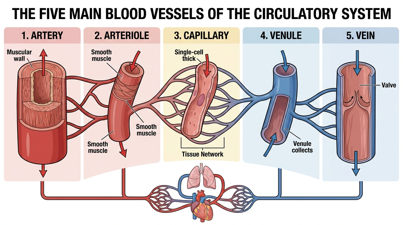
Blood vessels are hollow structures that form a connected network throughout the body. They include arteries, arterioles, capillaries, venules, and veins. Arteries generally carry blood away from the heart, while veins return blood toward the heart. Capillaries connect the arterial and venous sides of the circulation and provide the principal location for exchange between blood and tissues.
At the heart, however, five vessels are particularly important for understanding the overall circulation:
- Aorta
- Superior vena cava
- Inferior vena cava
- Pulmonary artery
- Pulmonary veins
Together, these vessels connect the heart with the systemic and pulmonary circulations.
2. The Two Circulatory Circuits
The five major vessels can be understood most easily by dividing circulation into two interconnected circuits.
2.1 Pulmonary circulation
Pulmonary circulation transports blood between the heart and lungs.
The sequence is:
Right ventricle → pulmonary arteries → lungs → pulmonary veins → left atrium
Blood leaving the right ventricle through the pulmonary arteries is relatively low in oxygen and relatively high in carbon dioxide. In the lungs, carbon dioxide is released and oxygen enters the blood. The oxygen-rich blood then returns to the heart through the pulmonary veins.
2.2 Systemic circulation
Systemic circulation transports blood between the heart and the rest of the body.
The simplified sequence is:
Left ventricle → aorta → arteries → capillaries → veins → venae cavae → right atrium
The left ventricle generates the pressure required to send oxygen-rich blood through the aorta and throughout the body. After oxygen and nutrients are delivered to tissues, blood returns toward the heart through progressively larger veins and ultimately through the superior and inferior venae cavae.
3. The Aorta
3.1 General description
The aorta is the principal artery of the systemic circulation and the largest artery in the human body.
It begins at the left ventricle of the heart. When the left ventricle contracts, oxygen-rich blood is ejected through the aortic valve into the aorta.
The aorta then distributes blood to arteries that supply virtually every major region of the body.
Simplified pathway
Left ventricle → aortic valve → aorta → systemic arteries → tissues
The aorta therefore represents the major departure point for oxygenated blood entering systemic circulation.
3.2 Major sections of the aorta
Anatomically, the aorta can be divided into several major portions:
- Ascending aorta
- Aortic arch
- Descending thoracic aorta
- Abdominal aorta
The ascending aorta arises from the heart.
The aortic arch curves over the heart and gives rise to major arteries supplying the head, neck, and upper limbs.
The descending aorta travels downward through the thorax and abdomen, giving off branches that supply organs and tissues.
3.3 Functions of the aorta
The aorta performs several critical functions.
Distribution
It distributes oxygen-rich blood to the systemic circulation.
Pressure management
Its elastic walls expand during ventricular contraction and recoil between heartbeats. This elastic behavior helps maintain relatively continuous blood flow despite the heart’s pulsatile pumping action.
Branching
The aorta serves as the parent vessel for numerous major arteries.
These branches eventually become smaller arteries, arterioles, and capillary networks.
4. The Superior Vena Cava
4.1 General description
The superior vena cava is one of the two great veins that return oxygen-poor blood from the systemic circulation to the heart.
It drains blood primarily from areas above the diaphragm, including the head, neck, upper limbs, and upper portions of the chest.
It empties into the right atrium.
Simplified pathway
Head and upper body → veins → superior vena cava → right atrium
The superior vena cava is therefore a major return pathway for systemic venous blood.
4.2 Function
The fundamental function of the superior vena cava is to return blood to the heart after the blood has circulated through tissues in the upper part of the body.
This blood has generally delivered much of its oxygen and collected carbon dioxide and metabolic waste.
Once it enters the right atrium, it joins blood returning through the inferior vena cava.
5. The Inferior Vena Cava
5.1 General description
The inferior vena cava is the second major vena cava.
It returns blood from most areas below the diaphragm to the right atrium.
Its drainage territory includes the abdomen, pelvis, and lower limbs.
Simplified pathway
Lower body → veins → inferior vena cava → right atrium
Together, the superior and inferior venae cavae provide the principal venous return from the systemic circulation.
5.2 Importance of the inferior vena cava
The lower parts of the body are relatively distant from the heart, and venous blood must travel back against gravity from the lower limbs.
The venous system therefore uses several mechanisms to assist venous return. Many veins, particularly in the arms and legs, contain valves that help prevent backward movement of blood. Muscle contractions can also assist the movement of venous blood toward the heart.
6. The Pulmonary Arteries
6.1 General description
The pulmonary arteries carry blood from the right ventricle toward the lungs.
This creates one of the most important exceptions to the simple rule that arteries carry oxygen-rich blood.
An artery is defined by its direction of flow away from the heart, not by its oxygen content.
Therefore:
- Pulmonary arteries carry blood away from the heart.
- Their blood is relatively low in oxygen.
- They transport blood to the lungs for oxygenation.
The pulmonary arteries divide into right and left pulmonary arteries, supplying the respective lungs.
6.2 The pulmonary circulation
After blood leaves the right ventricle through the pulmonary artery, it travels through increasingly smaller vessels within the lungs.
Eventually it reaches capillary networks surrounding the alveoli.
At these microscopic exchange surfaces:
- Carbon dioxide moves from blood into the air within the alveoli.
- Oxygen moves from inhaled air into the blood.
The oxygenated blood then enters the pulmonary veins.
7. The Pulmonary Veins
7.1 General description
The pulmonary veins return oxygen-rich blood from the lungs to the heart.
They empty into the left atrium.
This makes them another important exception to a common oversimplification:
- Veins carry blood toward the heart.
- Pulmonary veins carry oxygen-rich blood.
Therefore, oxygen content does not determine whether a vessel is an artery or vein. Direction of blood flow does.
Simplified pathway
Lungs → pulmonary veins → left atrium
From the left atrium, blood passes through the mitral valve into the left ventricle, which then pumps it into the aorta.
8. The Five Vessels as One Continuous System
The five vessels should not be studied independently. They are components of one continuous circulation.
A simplified complete pathway is:
Body tissues
↓
Superior vena cava + inferior vena cava
↓
Right atrium
↓
Right ventricle
↓
Pulmonary arteries
↓
Lungs
↓
Pulmonary veins
↓
Left atrium
↓
Left ventricle
↓
Aorta
↓
Body tissues
This cycle repeats continuously throughout life.
9. Oxygen-Rich and Oxygen-Poor Blood
One of the most important principles in understanding circulation is that oxygen content and vessel classification are not the same thing.
| Vessel | Direction | Typical oxygen status |
|---|---|---|
| Aorta | Away from heart | Oxygen-rich |
| Superior vena cava | Toward heart | Oxygen-poor |
| Inferior vena cava | Toward heart | Oxygen-poor |
| Pulmonary arteries | Away from heart | Oxygen-poor |
| Pulmonary veins | Toward heart | Oxygen-rich |
The pulmonary arteries and pulmonary veins are particularly important because they demonstrate why the terms “artery” and “vein” describe direction of blood flow, rather than simply whether blood contains oxygen.
10. Relationship With the Four Chambers of the Heart
The five major vessels connect directly or indirectly with the four chambers of the heart.
Right atrium
Receives systemic venous blood from:
- Superior vena cava
- Inferior vena cava
Right ventricle
Receives blood from the right atrium and pumps it toward the lungs through the pulmonary arteries.
Left atrium
Receives oxygen-rich blood from the pulmonary veins.
Left ventricle
Receives blood from the left atrium and pumps it into the aorta.
This arrangement creates a highly organized two-sided pump.
11. The Role of Heart Valves
Blood must move in a controlled direction.
The heart contains four valves that help prevent backward flow:
- Tricuspid valve
- Pulmonary valve
- Mitral valve
- Aortic valve
The pulmonary valve controls flow between the right ventricle and pulmonary artery, while the aortic valve controls flow between the left ventricle and aorta.
The valves therefore work together with the major blood vessels to establish directional circulation.
12. Arteries, Veins and Capillaries
Although this thesis focuses on five major vessels, these vessels are only the large-scale components of a much larger vascular network.
12.1 Arteries
Arteries carry blood away from the heart.
Their walls are generally thick and muscular because they experience relatively high pressure. They branch into progressively smaller arteries and arterioles.
12.2 Capillaries
Capillaries are microscopic vessels.
Their extremely thin walls permit exchange between blood and tissues.
Oxygen and nutrients can move from blood into tissues, while carbon dioxide and other waste products move in the opposite direction.
12.3 Veins
Veins carry blood toward the heart.
They generally operate under lower pressure than arteries and have thinner walls. Some veins contain valves that assist one-way blood movement, especially in the limbs.
13. Why Blood Vessels Are Essential
Blood vessels perform much more than simple transportation.
They enable the body to:
- Deliver oxygen
- Deliver nutrients
- Remove carbon dioxide
- Remove metabolic waste
- Transport hormones
- Distribute heat
- Support immune responses
- Maintain tissue oxygenation
- Help regulate blood pressure
- Maintain internal physiological balance
The cardiovascular system therefore functions as an integrated transportation and regulation network.
14. Blood Pressure and Vessel Function
Blood pressure is generated largely by the pumping action of the heart and influenced by the resistance and elasticity of blood vessels.
Arteries must withstand higher pressures than veins.
Arterial walls contain substantial elastic and muscular components that allow them to expand and contract in response to changes in blood pressure. Arterioles play an especially important role in regulating how much blood reaches particular tissues.
Veins, by contrast, operate at much lower pressures and can serve as important reservoirs for blood volume.
15. The Circulatory System as a Transportation Network
A useful way to understand the five vessels is to compare circulation with a transportation system.
The aorta resembles the major highway leaving a central distribution station.
The arterial branches resemble smaller roads distributing supplies.
The capillaries resemble local delivery points where materials are exchanged.
The veins resemble return routes.
The superior and inferior venae cavae are major return channels.
The pulmonary arteries and pulmonary veins form a specialized transportation route between the heart and lungs.
The heart functions as the pump that keeps the entire system moving.
16. Major Diseases Affecting Blood Vessels
Blood vessels can be affected by numerous diseases and disorders.
16.1 Atherosclerosis
Atherosclerosis involves the development of fatty and other deposits within artery walls. These deposits can narrow arteries and interfere with blood flow.
16.2 Aneurysm
An aneurysm is an abnormal enlargement or bulging of a weakened portion of a blood vessel.
The aorta is one of the major vessels in which aneurysms can occur.
16.3 Blood clots
Clots can obstruct vessels.
A clot in a vein may interfere with venous return, while a clot affecting an artery can reduce blood supply to tissues.
16.4 Varicose veins
When valves in veins do not function properly, blood can flow backward and veins may become enlarged and twisted. This is particularly common in superficial veins of the legs.
16.5 Vasculitis
Vasculitis involves inflammation of blood vessels and can affect their normal structure and function.
These conditions demonstrate why maintaining healthy blood vessels is fundamental to cardiovascular health.
17. The Five Vessels and the Human Body’s Oxygen Economy
The body has a continuous oxygen requirement.
The sequence can be summarized as follows:
1. Pulmonary arteries
Carry oxygen-poor blood to the lungs.
2. Lungs
Exchange carbon dioxide for oxygen.
3. Pulmonary veins
Return oxygen-rich blood to the left atrium.
4. Aorta
Distributes oxygen-rich blood to the body.
5. Superior and inferior venae cavae
Return oxygen-poor blood to the right atrium.
This cycle repeats continuously.
18. Why the System Is Called a Double Circulation
Humans possess a double circulatory system because blood passes through two major circuits.
Pulmonary circuit
Heart → lungs → heart
Systemic circuit
Heart → body → heart
The right side of the heart primarily drives pulmonary circulation, while the left side drives systemic circulation.
This separation allows the lungs to oxygenate blood before that blood is pumped at appropriate pressure to the rest of the body.
19. The Importance of Capillaries Between the Major Vessels
The five major vessels cannot perform their functions alone.
Between large arteries and large veins is an immense network of smaller vessels.
The progression is approximately:
Aorta → arteries → arterioles → capillaries → venules → veins → venae cavae
At the same time, the pulmonary circulation follows:
Pulmonary artery → smaller pulmonary arteries → arterioles → pulmonary capillaries → venules → pulmonary veins
The capillaries are therefore the crucial exchange interface between circulation and tissues.
20. A Simplified Master Model
The entire circulation can be remembered through five questions:
Where does systemic blood leave the heart?
Through the aorta.
Where does systemic blood return from the upper body?
Through the superior vena cava.
Where does systemic blood return from the lower body?
Through the inferior vena cava.
Where does oxygen-poor blood go after leaving the right side of the heart?
Through the pulmonary arteries to the lungs.
Where does oxygen-rich blood return from the lungs?
Through the pulmonary veins to the left atrium.
These five answers provide a powerful framework for understanding the cardiovascular system.
21. Clinical Importance
Understanding the five major vessels is fundamental to medical anatomy because abnormalities in these vessels can have consequences throughout the body.
The aorta is essential for systemic arterial distribution.
The venae cavae are essential for systemic venous return.
The pulmonary arteries are essential for transporting blood to the lungs.
The pulmonary veins are essential for returning oxygenated blood to the left side of the heart.
Because these vessels are central pathways, obstruction, abnormal enlargement, damage, or other disease can interfere with normal circulation.
22. Conclusion
The circulatory system is one of the body’s most sophisticated biological transportation networks. At its center, five major vessel groups provide the principal pathways connecting the heart with the body and lungs:
the aorta, superior vena cava, inferior vena cava, pulmonary arteries, and pulmonary veins.
The aorta carries blood from the left ventricle into systemic circulation.
The superior vena cava returns blood from the upper body.
The inferior vena cava returns blood from the lower body.
The pulmonary arteries carry oxygen-poor blood from the right side of the heart to the lungs.
The pulmonary veins return oxygen-rich blood from the lungs to the left side of the heart.
Together, these vessels form a continuous circulation that supports oxygen delivery, nutrient distribution, waste removal, temperature regulation, hormonal transport, and the maintenance of life.
The most important conceptual lesson is that arteries carry blood away from the heart and veins carry blood toward the heart. Oxygen content is a separate characteristic. This explains why the pulmonary arteries carry oxygen-poor blood while the pulmonary veins carry oxygen-rich blood.
Ultimately, the five major vessels should not be viewed as isolated tubes. They are components of one integrated cardiovascular network in which the heart, lungs, arteries, capillaries, and veins operate continuously as a coordinated system.
Key Terms
Aorta: The principal artery carrying blood from the left ventricle to systemic circulation.
Superior vena cava: Major vein returning blood from the upper body to the right atrium.
Inferior vena cava: Major vein returning blood from the lower body to the right atrium.
Pulmonary arteries: Vessels carrying oxygen-poor blood from the right ventricle to the lungs.
Pulmonary veins: Vessels carrying oxygen-rich blood from the lungs to the left atrium.
Capillaries: Microscopic vessels where blood-tissue exchange occurs.
Systemic circulation: Circulation between the heart and the rest of the body.
Pulmonary circulation: Circulation between the heart and lungs.
Artery: A blood vessel carrying blood away from the heart.
Vein: A blood vessel carrying blood toward the heart.
Selected References
- National Heart, Lung, and Blood Institute (NHLBI), How the Heart Works: How Blood Flows Through the Heart.
- National Heart, Lung, and Blood Institute (NHLBI), How the Lungs Work: The Respiratory System.
- MSD Manual, Biology of the Blood Vessels.
- MSD Manual, Overview of the Venous System.
- National Heart, Lung, and Blood Institute, Your Heart, Your Life educational materials.
`




Be First to Comment