
Abstract
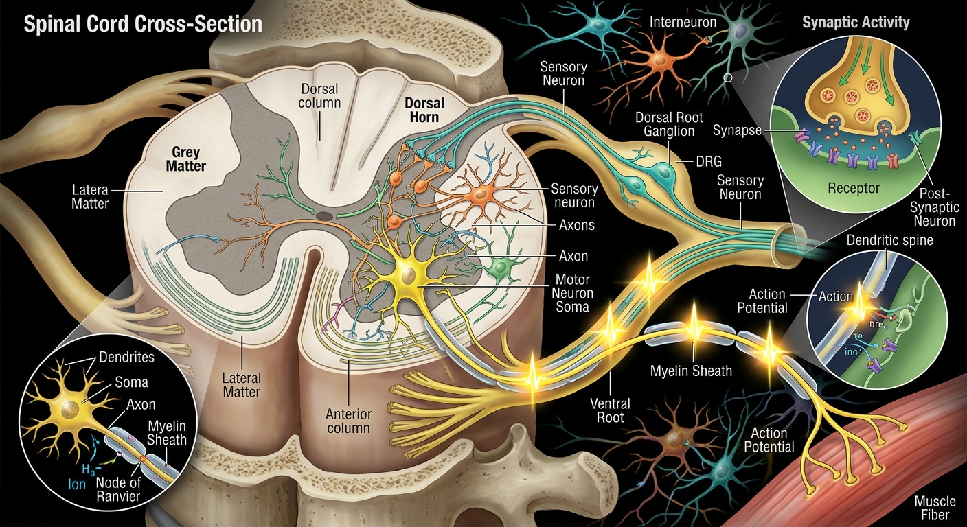
The human spinal cord is a highly organized biological communication system connecting the brain with the rest of the body. It is not simply a cable containing neurons. It is a living network containing neurons, glial cells, synapses, axons, blood vessels, protective membranes, and specialized circuits that continuously receive, process, and transmit information.
An important clarification is that adult neurons generally do not move up and down the spinal cord like electrical signals. What moves over long distances is primarily the electrical and chemical information carried along neuronal axons. The neurons themselves are mostly fixed in anatomical locations. During development, however, immature neurons and their precursor cells migrate to their appropriate positions.
This thesis explains the spinal cord from its large-scale anatomy down to the movement of signals through individual neurons.
Chapter 1 — What Is the Spinal Cord?
The spinal cord is part of the central nervous system (CNS), together with the brain.
It forms a continuous communication pathway between:
Brain ↔ Spinal cord ↔ Peripheral nerves ↔ Muscles, organs and sensory receptors
In an adult, the spinal cord is roughly cylindrical and extends from the lower brainstem through the vertebral canal. It is protected by:
- Vertebrae
- Meninges
- Cerebrospinal fluid
- Blood vessels
- Specialized cellular barriers
Its fundamental functions are:
- transmitting sensory information toward the brain;
- transmitting motor commands toward the body;
- coordinating reflexes;
- integrating local information;
- participating in autonomic regulation;
- generating and coordinating certain rhythmic motor patterns.
Chapter 2 — The Basic Architecture
A useful way to understand the spinal cord is to imagine it as a multi-layer biological computing system.
                 BRAIN
                   │
                   │
          ┌────────▼────────┐
          │  Spinal cord    │
          │                 │
Sensory ──►  INFORMATION    ├──► Motor
input     │  PROCESSING     │    output
          │                 │
          └─────────────────┘
             │    │    │
             ▼    ▼    ▼
           Skin  Organs Muscles
But the actual architecture is much more sophisticated.
At the microscopic level, the spinal cord contains two major tissue arrangements:
Gray matter
Contains many:
- neuron cell bodies;
- dendrites;
- synapses;
- local neuronal circuits;
- glial cells.
White matter
Contains large numbers of myelinated axons organized into pathways.
A simplified cross-section looks like:
              Dorsal / posterior
                     ↑
          ┌─────────────────────┐
          │   WHITE MATTER      │
          │ ┌─────────────────┐ │
          │ │   GRAY MATTER   │ │
          │ │     ╲   ╱       │ │
          │ │      ╲ ╱        │ │
          │ │       ●         │ │
          │ │      ╱ ╲        │ │
          │ └─────────────────┘ │
          │   WHITE MATTER      │
          └─────────────────────┘
                     ↓
              Ventral / anterior
The gray matter has a characteristic butterfly or H-shaped appearance in cross-section.
Chapter 3 — The Neuron: The Fundamental Processing Unit
A neuron is a specialized cell designed to receive, process and transmit information.
Its major components are:
       Dendrites
       ↓ ↓ ↓
     \  | |  /
      \ | | /
      [CELL BODY]
          │
          │ Axon
          │
     == == == == == ==>
          │
          ▼
       Axon terminal
          │
       Synapse
3.1 Dendrites
Dendrites receive information from other cells.
They may receive:
- excitatory signals;
- inhibitory signals;
- modulatory signals.
3.2 Cell body
The soma contains the nucleus and much of the machinery required to keep the neuron alive.
It integrates incoming information.
3.3 Axon
The axon is a specialized projection that can carry electrical signals over considerable distances.
Some spinal axons are extremely long relative to the size of the cell body.
3.4 Axon terminal
The terminal communicates with another neuron, muscle cell or glandular cell.
Communication commonly occurs through neurotransmitters released into the synaptic cleft.
Chapter 4 — Do Neurons Actually Move Through the Spinal Cord?
This is one of the most important concepts.
Adult nervous system
Most mature neurons are stationary.
For example, a motor neuron located in the spinal cord does not normally travel from the lower spinal cord to the brain every time you move your hand.
Instead:
The neuron stays in place while electrical information travels along its axon.
Think of it like a fixed computer connected to a network cable.
The computer remains stationary.
Information moves through the network.
Similarly:
Neuron cell body
      │
      │
      ▼
   AXON
      │
      │ electrical signal
      ▼
Axon terminal
      │
      ▼
   Synapse
Therefore, it is more accurate to speak about the movement of neural information rather than the physical movement of mature neurons.
Chapter 5 — What Actually Moves?
Several different processes can be described as “movement” in neuroscience.
5.1 Electrical signals
The action potential propagates along the axon.
5.2 Ions
Charged particles such as:
- Na⁺
- K⁺
- Ca²⁺
- Cl⁻
move through cellular membranes or participate in electrical gradients.
5.3 Neurotransmitters
Chemical messengers cross synaptic gaps.
5.4 Molecular cargo
Proteins, vesicles and other materials are transported inside axons.
5.5 Information
Patterns of neuronal activity travel through networks.
These are fundamentally different types of movement.
Chapter 6 — Electrical Architecture of a Neuron
The neuron maintains an electrical difference across its membrane.
The resting membrane potential results from several factors, including:
- ion concentration gradients;
- membrane permeability;
- ion channels;
- the sodium-potassium pump.
A simplified representation is:
Outside neuron
   Na+  Na+  Na+
────────────────────
    Cell membrane
────────────────────
   K+   K+   K+
Inside neuron
The membrane separates different ionic environments.
When sufficient excitatory input reaches a neuron, voltage-sensitive channels can produce an action potential.
Chapter 7 — The Action Potential
The action potential is the major mechanism for rapid long-distance electrical signaling in neurons.
Simplified sequence:
REST
 │
 ▼
Threshold reached
 │
 ▼
Depolarization
 │
 ▼
Repolarization
 │
 ▼
Hyperpolarization
 │
 ▼
Return toward resting state
During depolarization, voltage-gated sodium channels open, allowing sodium ions to enter.
Then sodium channels become inactivated and potassium channels contribute to repolarization.
The important point is:
The action potential propagates along the axon rather than the neuron itself travelling through the spinal cord.
Chapter 8 — Myelin: The High-Speed Insulation System
Many spinal axons are surrounded by myelin.
In the CNS, myelin is produced by oligodendrocytes.
Myelin electrically insulates portions of the axon.
Between myelinated sections are small gaps called:
Nodes of Ranvier
The arrangement can be simplified as:
Axon:

====[MYELIN]====●====[MYELIN]====●====[MYELIN]====

                ↑                 ↑
             Node              Node
The action potential is regenerated at successive nodes.
This allows rapid conduction through a process called saltatory conduction.
Chapter 9 — The Spinal Cord as a Two-Way Information Highway
There are two broad directions of information flow.
Ascending pathways
Carry sensory information toward the brain.
BODY
 │
 │ sensory information
 ▼
SPINAL CORD
 │
 ▼
BRAIN
Descending pathways
Carry commands from the brain toward the spinal cord and body.
BRAIN
 │
 │ motor commands
 ▼
SPINAL CORD
 │
 ▼
MOTOR NEURONS
 │
 ▼
MUSCLES
However, these pathways are not simply two wires.
They contain many parallel pathways with different functions.
Chapter 10 — Sensory Neurons
Sensory neurons detect information from the body and environment.
They can provide information about:
- touch;
- pressure;
- vibration;
- temperature;
- pain;
- muscle length;
- joint position;
- internal organ states.
Many sensory neuron cell bodies associated with spinal nerves are located in the dorsal root ganglia.
A simplified pathway is:
Sensory receptor
      │
      ▼
Sensory neuron
      │
      ▼
Dorsal root
      │
      ▼
Spinal cord
      │
      ├──► Local reflex circuit
      │
      └──► Ascending pathway
                  │
                  ▼
                 Brain
Chapter 11 — Motor Neurons
Motor neurons transmit commands to muscles.
Their cell bodies for skeletal muscle control are located primarily in the ventral horn of spinal gray matter.
Simplified:
Brain
 │
 ▼
Descending pathway
 │
 ▼
Spinal interneuron / motor circuit
 │
 ▼
Motor neuron
 │
 ▼
Peripheral nerve
 │
 ▼
Neuromuscular junction
 │
 ▼
Skeletal muscle
The motor neuron itself does not travel from the brain to the muscle.
Its axon extends from the spinal cord into the peripheral nerve and ultimately reaches the muscle.
Chapter 12 — Interneurons: The Internal Computing Network
Interneurons are crucial to spinal-cord processing.
They connect neurons within the nervous system.
They can:
- connect sensory neurons to motor neurons;
- inhibit other neurons;
- amplify signals;
- coordinate opposing muscles;
- participate in reflexes;
- contribute to locomotor circuits.
This means the spinal cord is not merely a transmission cable.
It is also a distributed biological computer.
Chapter 13 — The Reflex Arc
One of the clearest examples of spinal information processing is the reflex.
Suppose a sensory receptor detects a sudden stimulus.
A simplified circuit can be represented as:
Stimulus
   │
   ▼
Sensory receptor
   │
   ▼
Sensory neuron
   │
   ▼
Spinal cord
   │
   ▼
Interneuron
   │
   ▼
Motor neuron
   │
   ▼
Muscle
   │
   ▼
Response
The brain may also receive information about the event.
This illustrates an important principle:
Some nervous-system responses can be organized within the spinal cord without waiting for conscious processing by the brain.
Chapter 14 — Gray Matter Architecture
The gray matter is divided broadly into regions known as horns.
Dorsal horn
Primarily associated with sensory processing.
Ventral horn
Contains motor neurons controlling skeletal muscles.
Lateral horn
Present prominently at certain spinal levels and associated with autonomic nervous-system circuitry.
Simplified:
             DORSAL
               ↑
          ┌───────────┐
          │   DORSAL  │
          │   HORN    │
          │     ╲ ╱   │
          │      X    │
          │     ╱ ╲   │
          │  VENTRAL  │
          │    HORN   │
          └───────────┘
               ↓
             VENTRAL
Chapter 15 — White Matter Architecture
White matter surrounds the gray matter.
It contains bundles of axons called tracts.
These pathways can travel:
- upward;
- downward;
- between spinal segments.
The major longitudinal divisions are often described as:
- dorsal funiculus;
- lateral funiculus;
- ventral funiculus.
These contain numerous specialized pathways.
Chapter 16 — Ascending Sensory Pathways
Different sensory modalities travel through different anatomical systems.
For example, information concerning fine touch, vibration and conscious proprioception is conveyed primarily through the dorsal column–medial lemniscus system.
Pain and temperature information are primarily associated with the anterolateral/spinothalamic system.
This organization allows the nervous system to preserve different kinds of information.
A simplified conceptual model:
                 BRAIN
                   ▲
                   │
       ┌───────────┼───────────┐
       │           │           │
    Touch       Vibration     Pain
       ▲           ▲           ▲
       │           │           │
       └──────── SPINAL CORD ──┘
                   ▲
                   │
                 BODY
The real pathways involve crossings, multiple neurons and multiple processing stations.
Chapter 17 — Descending Motor Pathways
The brain sends motor information downward through several pathways.
The best-known voluntary motor pathway is the corticospinal tract.
A simplified organization is:
Motor cortex
     │
     ▼
Brainstem
     │
     ▼
Spinal cord
     │
     ▼
Interneurons
     │
     ▼
Motor neurons
     │
     ▼
Muscles
The corticospinal system is particularly important for skilled voluntary movement.
Chapter 18 — The Spinal Cord Is Segmented
The spinal cord has a segmental organization associated with spinal nerves.
The commonly described spinal levels are:
- cervical;
- thoracic;
- lumbar;
- sacral;
- coccygeal.
There are 31 pairs of spinal nerves.
They connect the CNS with different regions of the body.
This resembles a distributed network:
                  BRAIN
                    │
             ┌──────┴──────┐
             │ SPINAL CORD │
             └──────┬──────┘
        ┌────────────┼────────────┐
        │            │            │
    Cervical      Thoracic     Lumbar/
    nerves        nerves       sacral nerves
        │            │            │
      Arms         Trunk       Legs/pelvis
Chapter 19 — Neuronal Communication Is Mostly Chemical at Synapses
When an action potential reaches an axon terminal, it can cause calcium channels to open.
Calcium enters the terminal and promotes neurotransmitter release.
Simplified:
Action potential
       │
       ▼
Axon terminal
       │
       ▼
Ca²⁺ entry
       │
       ▼
Neurotransmitter release
       │
       ▼
Synaptic cleft
       │
       ▼
Receptors on next cell
The receiving neuron then changes its electrical state.
Thus neural communication combines:
Electrical signaling + chemical signaling + electrical signaling
Chapter 20 — Excitation and Inhibition
The spinal cord must balance neuronal activity.
Two fundamental influences are:
Excitation
Makes a neuron more likely to generate an action potential.
Inhibition
Makes it less likely.
This is essential for coordinated movement.
For example, when one muscle group contracts, the nervous system can simultaneously inhibit opposing muscles through specialized spinal circuits.
This principle contributes to smooth movement.
Chapter 21 — Neural Information Is Not a Single Electrical “Current”
A common misconception is that the nervous system works like a copper electrical wire.
It does not.
A spinal neural signal involves:
- Membrane voltage changes.
- Ion-channel activity.
- Action-potential propagation.
- Neurotransmitter release.
- Receptor activation.
- Network integration.
- New patterns of neuronal activity.
Therefore:
The nervous system is an electrochemical information-processing network, not simply an electrical cable.
Chapter 22 — How Fast Does Information Travel?
Neural conduction speed varies considerably.
Factors include:
- axon diameter;
- myelination;
- membrane properties;
- temperature;
- axonal structure.
Some heavily myelinated axons conduct signals at well over 100 m/s, whereas smaller unmyelinated fibers conduct much more slowly.
Thus there is no single “speed of the spinal cord.”
Different information channels operate at different conduction speeds.
Chapter 23 — Axonal Transport: Another Kind of Movement
There is another important meaning of movement within neurons.
The neuron transports molecular materials along its axon.
This is called axonal transport.
Two broad directions exist:
Anterograde transport
Cell body ─────────► Axon terminalRetrograde transport
Axon terminal ─────► Cell bodyMolecular cargo can include:
- proteins;
- membrane components;
- vesicles;
- signaling molecules;
- damaged cellular material destined for processing.
This movement is much slower than an action potential.
Motor proteins such as kinesins and dyneins move cargo along microtubules.
Chapter 24 — The Cytoskeleton: Internal Railway System
Inside neurons are structural elements including:
- microtubules;
- neurofilaments;
- actin filaments.
Microtubules provide tracks for intracellular transport.
A conceptual analogy:
Neuron cell body
      │
      │ microtubule tracks
      ═══════════════════►
      │       cargo
      │       ●
      │
      ▼
Axon terminal
This is molecular transportation inside the neuron, not movement of the neuron itself.
Chapter 25 — Glial Cells: The Supporting Infrastructure
Neurons cannot operate alone.
Important CNS glial cells include:
Astrocytes
They participate in:
- metabolic support;
- ion regulation;
- neurotransmitter regulation;
- synaptic environments;
- blood–brain/spinal barriers.
Oligodendrocytes
Produce myelin around CNS axons.
Microglia
Serve important immune and surveillance functions in the CNS.
Ependymal cells
Line the ventricular and central-canal system and contribute to the specialized environment associated with cerebrospinal fluid.
Therefore, the spinal cord is an ecosystem of interacting cell types.
Chapter 26 — Blood Supply
Neurons have enormous metabolic requirements.
They require continuous supplies of:
- oxygen;
- glucose;
- other metabolic substrates.
The spinal cord receives blood through a network involving longitudinal arteries and reinforcing segmental vessels.
If blood flow is severely interrupted, nervous tissue can be damaged because neurons have limited tolerance for prolonged oxygen deprivation.
Chapter 27 — Protection of the Spinal Cord
The spinal cord is protected by three major meninges:
- Dura mater
- Arachnoid mater
- Pia mater
Cerebrospinal fluid also contributes to protection and the physiological environment.
The vertebral column provides mechanical protection.
Thus the architecture is:
Vertebra
   │
   ▼
Dura
   │
   ▼
Arachnoid
   │
   ▼
CSF
   │
   ▼
Pia
   │
   ▼
Spinal cord
Chapter 28 — Neuron Development: When Neurons Actually Move
There is an important distinction between the adult spinal cord and the developing nervous system.
During embryonic development, neural precursor cells are generated and then undergo carefully regulated processes involving:
- proliferation;
- migration;
- differentiation;
- axon growth;
- synapse formation;
- programmed cell death;
- circuit refinement.
So, during development, cells can physically migrate.
The developing spinal cord begins from the neural tube.
Different populations of cells become organized into dorsal and ventral functional domains.
Very broadly:
Developing neural tube

        Dorsal
          ↑
   Sensory-related
      domains
          │
          │
      Central
       region
          │
          │
   Motor-related
      domains
          ↓
        Ventral
This developmental organization establishes much of the architecture seen in the mature spinal cord.
Chapter 29 — Axon Guidance
Developing neurons must send axons to appropriate targets.
Growing axons possess specialized structures called growth cones.
The growth cone explores its environment and responds to molecular guidance signals.
Conceptually:
Growing axon
───────────────►
              (growth cone)
                   ◉
                 / | \
                /  |  \
      environmental guidance cues
Molecular signals can attract or repel developing axons.
This allows enormous numbers of neurons to establish organized connections.
Chapter 30 — Synaptic Plasticity
The spinal cord is not a rigid circuit.
Connections can change their strength.
This property is called neural plasticity.
Plasticity can involve:
- changes in receptor numbers;
- changes in neurotransmitter release;
- alterations in synaptic strength;
- structural changes in synapses;
- changes in neuronal excitability;
- changes in network organization.
Therefore:
The spinal cord is structurally organized but functionally adaptable.
Chapter 31 — The Spinal Cord as a Distributed Computer
A useful conceptual model is:
                    BRAIN
                      │
            ┌─────────┴─────────┐
            │                   │
       Descending            Ascending
       information           information
            │                   ▲
            ▼                   │
       ┌─────────────────────────────┐
       │       SPINAL CORD            │
       │                              │
       │  Sensory circuits            │
       │       ↓                      │
       │  Interneurons                │
       │       ↓                      │
       │  Motor circuits              │
       │                              │
       │  Reflex circuits             │
       │                              │
       │  Autonomic circuits          │
       └─────────────────────────────┘
            │                   ▲
            ▼                   │
         MUSCLES              SENSORS
It performs several computational operations:
Input → integration → transformation → output → feedback
This is one reason why simply describing the spinal cord as a “wire” is inadequate.
Chapter 32 — A Complete Movement Example
Consider voluntarily moving your hand.
A simplified chain is:
Step 1 — Intention
Brain networks generate a motor command.
Step 2 — Descending transmission
Signals travel through descending pathways.
Step 3 — Spinal processing
The descending information interacts with spinal interneurons and motor circuits.
Step 4 — Motor neuron activation
A motor neuron generates action potentials.
Step 5 — Peripheral nerve
The action potentials travel along the motor axon.
Step 6 — Neuromuscular junction
The motor neuron communicates with muscle fibers.
Step 7 — Muscle contraction
The muscle generates force.
Step 8 — Sensory feedback
Receptors provide information about:
- muscle length;
- tension;
- joint position;
- touch.
Step 9 — Feedback to CNS
Sensory information travels back toward the spinal cord and brain.
This produces a continuous control loop:
             BRAIN
            ↙     ↖
       command   feedback
          ↓         ↑
       SPINAL CORD
          ↓         ↑
     MOTOR SYSTEM  SENSORY SYSTEM
          ↓         ↑
        MUSCLE ─────┘
Chapter 33 — The Deep Principle: Movement of Information
The most useful distinction is therefore:
| Component | Does it move? | What happens? |
|---|---|---|
| Mature neuron | Usually no | Remains anatomically positioned |
| Action potential | Yes | Propagates along axon |
| Ions | Yes | Move through channels/membranes |
| Neurotransmitters | Yes | Cross synaptic spaces |
| Axonal cargo | Yes | Transported inside axons |
| Developing neurons | Yes | Some migrate during development |
| Sensory information | Yes | Propagates through neural pathways |
| Motor commands | Yes | Descend through neural pathways |
Chapter 34 — The Spinal Cord as a Biological Network
The architecture can ultimately be understood at five levels:
Level 1 — Molecular
Ion channels, receptors, neurotransmitters and proteins.
Level 2 — Cellular
Neurons and glial cells.
Level 3 — Synaptic
Connections between individual cells.
Level 4 — Circuit
Groups of neurons performing specific functions.
Level 5 — Systems
Ascending and descending pathways connecting the spinal cord with the brain and body.
MOLECULES
    ↓
CELLS
    ↓
SYNAPSES
    ↓
CIRCUITS
    ↓
PATHWAYS
    ↓
WHOLE NERVOUS SYSTEM
This hierarchy is fundamental to understanding neuroscience.
Chapter 35 — Final Scientific Perspective
The human spinal cord is best understood as a highly organized electrochemical information-processing network.
Its architecture contains:
- gray matter for extensive local neuronal processing;
- white matter containing long-distance axonal pathways;
- sensory neurons bringing information inward;
- motor neurons sending commands outward;
- interneurons creating local computational circuits;
- glial cells maintaining the neural environment;
- myelin increasing conduction efficiency;
- synapses enabling cell-to-cell communication;
- vascular networks supporting enormous metabolic requirements.
The mature neurons themselves generally remain in their anatomical locations. What travels through the spinal system is principally electrical activity, chemical signaling and molecular cargo.
The deepest concept is therefore:
The spinal cord does not primarily transport neurons; it transports and transforms information through networks of neurons.
This makes the spinal cord simultaneously a communication system, biological computer, reflex controller, sensory processor and motor-control network.
And unlike a conventional computer, its computation is performed by living cells whose connections, chemistry and electrical properties can continuously adapt.




Be First to Comment