
The human brain stands as one of nature’s most extraordinary achievements—a three-pound organ containing a universe of complexity that enables consciousness, thought, emotion, and every aspect of human experience. Understanding the cellular composition, neural connectivity, and vascular support of this remarkable organ reveals the intricate biological machinery underlying our mental lives.
The Cellular Census: Neurons and Beyond
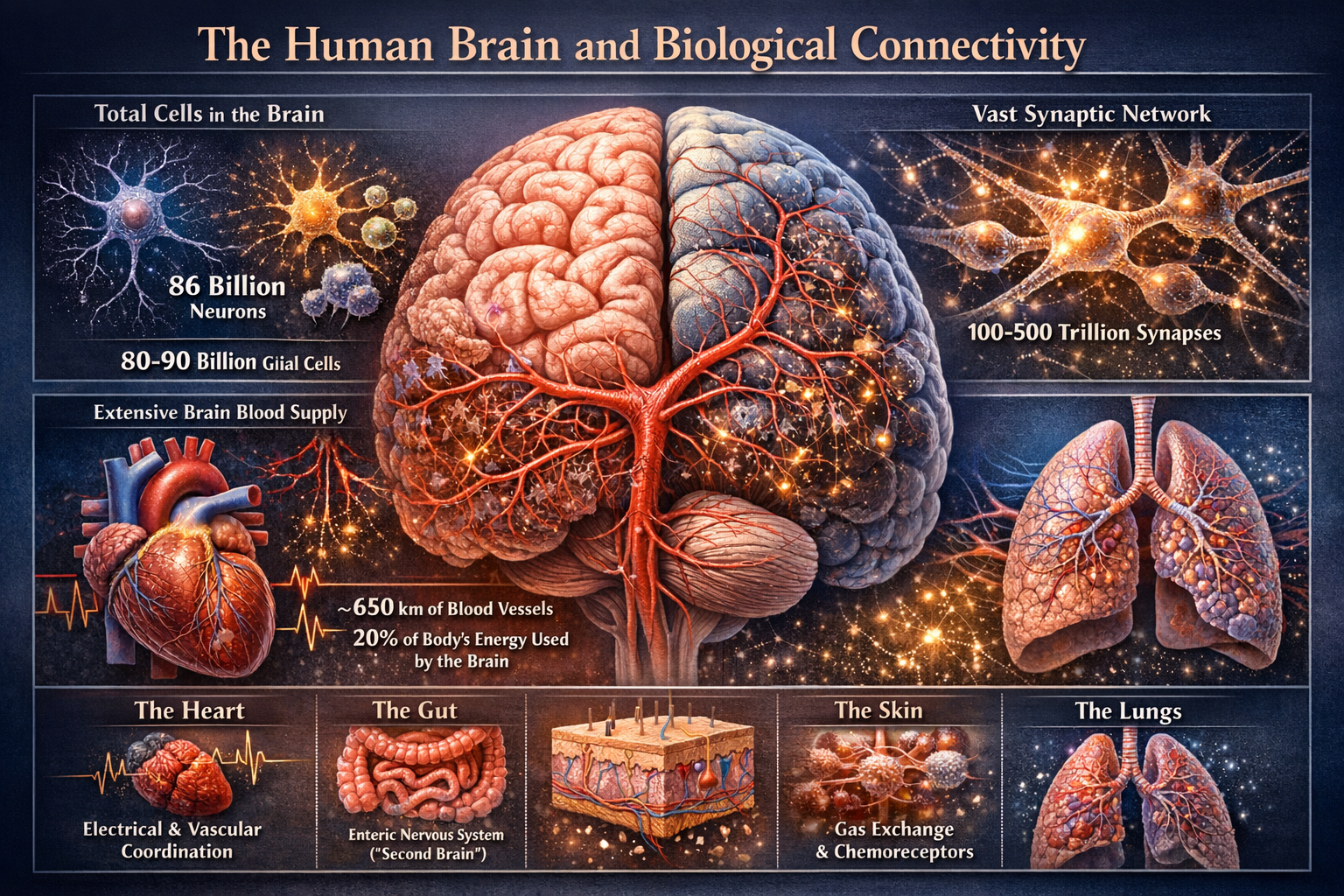
For decades, the commonly cited figure of 100 billion neurons in the human brain dominated scientific literature and popular understanding. However, more recent and rigorous methodological approaches have refined this estimate considerably. Using a technique called the isotropic fractionator method, neuroscientist Suzana Herculano-Houzel and colleagues determined that the adult human brain contains approximately 86 billion neurons—a substantial revision, though still representing an astronomical number of cellular units dedicated to information processing.
Yet neurons tell only part of the story. The brain’s cellular landscape includes an equally impressive population of glial cells, which were historically dismissed as mere support structures but are now recognized as active participants in brain function. The human brain contains roughly 85 billion glial cells, bringing the total cell count to approximately 171 billion cells working in concert. These glial cells include astrocytes, which regulate the chemical environment around neurons and help form the blood-brain barrier; oligodendrocytes, which produce the myelin sheaths that insulate neuronal axons and dramatically increase signal transmission speed; and microglia, the brain’s resident immune cells that prune synapses and respond to injury or disease.
Regional Distribution and Specialization
The distribution of these neurons across brain regions reveals much about functional organization. The cerebellum, despite occupying only about 10 percent of the brain’s volume, houses approximately 69 billion neurons—roughly 80 percent of all the brain’s neurons. This dense packing reflects the cerebellum’s crucial roles in motor coordination, balance, fine motor control, and increasingly recognized contributions to cognitive functions including attention, language, and emotional regulation.
The cerebral cortex, the wrinkled outer layer responsible for higher-order thinking, consciousness, and complex cognitive processes, contains about 16 billion neurons. Though fewer in absolute number than the cerebellum, cortical neurons exhibit extraordinary connectivity and diversity, with each neuron potentially forming thousands of synaptic connections with other neurons. This elaborate networking enables the emergence of abstract thought, planning, decision-making, and self-awareness.
The remaining brain structures—including the basal ganglia involved in movement control and habit formation, the hippocampus essential for memory formation, the amygdala critical for emotional processing, and various brainstem nuclei controlling vital functions like breathing and heart rate—collectively house the remaining billion or so neurons.
The Synaptic Universe: Connectivity and Communication
If the number of neurons is impressive, the connectivity between them is almost incomprehensible. Each neuron can form synapses with thousands of other neurons, creating an estimated 100 trillion to 1,000 trillion synaptic connections throughout the brain. These synapses represent the functional units of neural communication, the points where electrical signals in one neuron trigger chemical neurotransmitter release that influences the electrical state of receiving neurons.
The cerebral cortex demonstrates the most elaborate connectivity patterns. A single cortical pyramidal neuron—the most common excitatory neuron type in the cortex—may receive input from 10,000 to 30,000 other neurons and project its output to thousands more. This creates dense local circuits within cortical columns and regions, while also establishing long-range connections that integrate information across distant brain areas.
The prefrontal cortex, located at the front of the brain, exemplifies this connectivity principle at the system level. This region maintains connections with virtually every other major brain region, including sensory cortices, motor areas, limbic structures involved in emotion, and memory systems. These widespread connections enable the prefrontal cortex to serve as an executive control center, integrating diverse information streams to guide goal-directed behavior, working memory, impulse control, and complex decision-making.
Association cortices—areas that integrate information from multiple sensory and cognitive domains rather than processing single modalities—also exhibit exceptionally rich connectivity. The parietal association cortex integrates spatial, sensory, and motor information for navigation and attention. The temporal association cortex links auditory, visual, and semantic information crucial for language comprehension and object recognition. These highly connected hubs serve as integration zones where disparate information converges to create unified, meaningful experiences.
The Brain’s Vascular Infrastructure: A Critical Support System
The brain’s extraordinary metabolic demands require an equally impressive vascular system. Despite representing only about 2 percent of body weight, the brain consumes approximately 20 percent of the body’s oxygen and glucose—a disproportionate energy allocation reflecting the costly nature of maintaining neural signaling and synaptic transmission.
The brain receives blood through four major arteries: the paired internal carotid arteries and the paired vertebral arteries, which merge to form the basilar artery. These vessels form the Circle of Willis at the brain’s base, an anatomical arrangement providing redundancy and collateral circulation to protect against vascular compromise. From these major vessels, arteries branch progressively into smaller arterioles and eventually into an extraordinarily dense capillary network.
The brain contains approximately 400 miles of blood vessels, creating a capillary network so dense that no neuron sits more than about 40 micrometers from a blood vessel. This intimate spatial relationship between neurons and vasculature ensures efficient oxygen and nutrient delivery while removing metabolic waste products. The blood-brain barrier, formed by specialized endothelial cells lining brain capillaries in conjunction with astrocytic end-feet, selectively controls which substances can pass from blood into brain tissue, protecting the delicate neural environment from potentially harmful agents while permitting essential nutrients.
Functionally active brain regions exhibit increased blood flow through a phenomenon called neurovascular coupling. When neurons increase their firing rates, local blood vessels dilate within seconds, increasing blood flow to meet heightened metabolic demands. This coupling between neural activity and blood flow forms the basis of functional MRI imaging, which detects these localized blood flow changes to map brain activity.
White Matter: The Brain’s Information Superhighways
While gray matter containing neuronal cell bodies captures much attention, white matter—composed of myelinated axonal tracts—occupies nearly half the brain’s volume and enables the long-distance communication essential for integrated brain function. The corpus callosum, the largest white matter tract containing approximately 200 to 250 million axonal fibers, connects the brain’s two hemispheres, enabling interhemispheric communication and coordination.
Other major white matter tracts include the corona radiata, which fans out from the internal capsule to connect cortical areas with subcortical structures; the arcuate fasciculus, linking language comprehension and production areas; and the cingulum bundle, connecting limbic system components involved in emotion and memory. These tracts create the structural connectivity that allows functionally specialized brain regions to work together as integrated networks.
The quality and integrity of white matter connections significantly influence cognitive performance. Myelination continues through adolescence and into the mid-twenties, particularly in prefrontal regions, corresponding with improvements in executive function and impulse control. Age-related white matter decline contributes to cognitive slowing and reduced processing efficiency in older adults.
Functional Networks and Their Purposes
Modern neuroscience increasingly emphasizes distributed networks rather than isolated regions as the fundamental units of brain function. Several key networks have been identified through functional imaging and connectivity analyses.
The default mode network activates when the mind is at rest or engaged in internal mentation—self-referential thought, autobiographical memory retrieval, imagining the future, and theory of mind. This network includes the medial prefrontal cortex, posterior cingulate cortex, precuneus, and lateral parietal regions. Its connectivity patterns suggest a role in constructing mental simulations and maintaining self-awareness.
The salience network, anchored by the anterior insula and anterior cingulate cortex, detects behaviorally relevant stimuli and coordinates switching between other networks. It maintains extensive connections with limbic structures and helps direct attention toward salient information while filtering less relevant inputs.
The central executive network, centered on the dorsolateral prefrontal cortex and posterior parietal cortex, supports working memory, attention control, and goal-directed cognition. This network exhibits extensive connectivity with sensory, motor, and memory systems, enabling flexible, adaptive behavior.
Sensory and motor networks show more localized, hierarchical organization, with primary sensory cortices receiving direct inputs from thalamic relay nuclei and progressively processing information through increasingly complex feature representations in higher-order areas. Motor systems similarly progress from primary motor cortex to premotor and supplementary motor areas involved in movement planning and sequencing.
The Thalamus: The Brain’s Central Hub
While discussions of connectivity often focus on cortical regions, the thalamus deserves special recognition as perhaps the brain’s most highly connected structure relative to its size. This centrally located structure serves as a relay station for virtually all sensory information (except olfaction) traveling to the cortex. It maintains reciprocal connections with essentially all cortical areas, and increasingly, research reveals its active role in regulating cortical activity, consciousness, and attention rather than serving as a mere passive relay.
The thalamus contains distinct nuclei specialized for different functions: the lateral geniculate nucleus processes visual information, the medial geniculate nucleus handles auditory signals, the ventral posterior nucleus conveys somatosensory information, and the ventral lateral and ventral anterior nuclei relay motor information from the cerebellum and basal ganglia. Higher-order thalamic nuclei, including the mediodorsal and pulvinar nuclei, maintain extensive cortical connections and contribute to attention, cognition, and cortical coordination.
Energy Metabolism and Neuronal Demand
The brain’s high connectivity translates directly into substantial energy requirements. Maintaining ionic gradients across neuronal membranes, synthesizing and packaging neurotransmitters, and supporting synaptic transmission all require ATP, the cellular energy currency. Glucose serves as the brain’s primary fuel, metabolized through glycolysis and oxidative phosphorylation to generate ATP.
Different brain regions and cell types exhibit varying metabolic rates. Gray matter, packed with cell bodies and synapses, consumes more energy than white matter. During intense cognitive effort, activated regions may increase their metabolic rate by 50 percent or more above baseline. The astounding fact remains that even at rest, the brain continuously consumes substantial energy maintaining its baseline activity—the spontaneous neural firing and synaptic activity that characterizes the brain’s intrinsic organization.
Conclusion: An Integrated Perspective
The human brain represents a masterpiece of biological organization—86 billion neurons supported by equal numbers of glial cells, interconnected through hundreds of trillions of synapses, sustained by hundreds of miles of blood vessels, and organized into functionally specialized yet highly integrated networks. No single region dominates in terms of pure connectivity; rather, different structures serve as hubs within specific networks while maintaining broader integration with the whole.
The cerebral cortex, particularly association areas and the prefrontal cortex, exhibits the most elaborate and diverse connectivity patterns, enabling the integration necessary for complex cognition. The thalamus serves as a central hub coordinating cortical activity. The cerebellum, while containing the most neurons, operates somewhat more independently with specialized computations for motor control and coordination.
This intricate architecture emerges through development, shaped by genetic programs and experiential sculpting, refined through learning and adaptation, and maintained through precisely regulated metabolic support. Understanding this architecture not only satisfies scientific curiosity but provides essential foundation for addressing neurological and psychiatric disorders, developing brain-computer interfaces, and ultimately comprehending the biological basis of human consciousness and cognition. The brain’s complexity reminds us that we are only beginning to understand the full richness of the organ that makes understanding itself possible.
Be First to Comment