
Abstract
Myelinated fibres are among the most important structural components of the human nervous system. They enable the brain to transmit electrical signals rapidly and efficiently between neurons, allowing humans to think, learn, remember, speak, coordinate movement, interpret sensory information, and respond to the environment.
The word myelinated refers to nerve fibres that are covered by a specialised insulating layer called the myelin sheath. This sheath is formed primarily by specialised glial cells and surrounds the axons of many neurons.
To understand myelinated fibres properly, we must examine their biological building blocks, microscopic architecture, electrical mechanisms, relationship with brain networks, role in learning, development throughout life, and significance in neurological health.


8
Chapter 1: What are myelinated fibres?
A myelinated fibre is a nerve fibre whose axon is surrounded by a myelin sheath.
A typical neuron contains three main structural parts:

1. Dendrites
These branched structures receive many incoming signals from other neurons.

2. Cell body (soma)
Contains the nucleus and much of the machinery that keeps the neuron alive. The neuron integrates incoming information and, when conditions are met, initiates an electrical impulse.

3. Axon
A long extension that carries electrical impulses away from the cell body toward other neurons, muscles, or glands. Many axons have a myelin sheath, although some do not.
What makes a fibre myelinated?
The defining feature is the myelin sheath: a multilayered membrane rich in lipids and proteins that wraps around the axon.
Think of an axon as an electrical communication cable. The axon carries the signal, while myelin acts somewhat like insulation around a cable. However, biological nerve fibres are more sophisticated than manufactured wires: they use ion movement across cell membranes to generate and regenerate electrical impulses.
The central principle: myelin helps electrical signals travel faster and with less current leaking across the axonal membrane.
Chapter 2: The microscopic architecture of a myelinated fibre
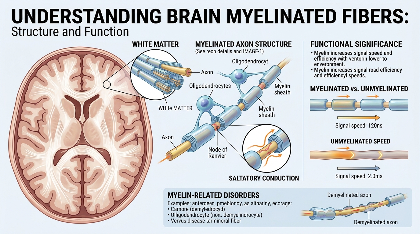
A myelinated fibre is not simply a neuron covered with insulation. It is a carefully organised biological communication system consisting of an axon, layers of myelin, specialised gaps, ion channels, and supporting glial cells.

The seven important structural elements
| Structure | Main function |
|---|---|
| 1. Axon | Carries electrical impulses. |
| 2. Myelin sheath | Insulates the axon and improves conduction efficiency. |
| 3. Internode | The long, myelin-covered segment between two nodes. |
| 4. Node of Ranvier | A gap where electrical impulses are regenerated. |
| 5. Paranode | Anchors and organises the ends of adjacent myelin segments. |
| 6. Juxtaparanode | A neighbouring region containing specialised potassium channels. |
| 7. Ion channels | Regulate sodium and potassium movement across the axonal membrane. |
These structures work together to maintain fast, reliable communication. Their organisation is essential for normal nerve conduction.
PMC
+1
2.1 The axon: the signal-carrying pathway
The axon is the central conducting component of a myelinated fibre.
Its membrane separates the fluid inside the axon from the fluid outside it. This separation allows electrical differences to develop across the membrane.
The membrane contains proteins called ion channels. Some open or close in response to changes in voltage, allowing charged particles such as sodium ions (\(\mathrm{Na^+}\)) and potassium ions (\(\mathrm{K^+}\)) to cross.
The movement of these ions produces the electrical signals used by the nervous system.
2.2 The myelin sheath: biological insulation
Myelin is produced by wrapping specialised cell membranes around an axon in multiple layers. These layers are rich in lipids and contain important proteins.
Its principal electrical effects are:
- Increased membrane resistance: less electrical current escapes across the covered membrane.
- Reduced membrane capacitance: less electrical charge is needed to change the voltage across the covered membrane.
- Improved conduction efficiency: electrical changes can spread rapidly along the axon toward the next node.
- Support for axonal health: myelin-forming cells also help supply and maintain the metabolic environment needed by axons.
Myelin therefore provides both electrical insulation and biological support.
NCBI Bookshelf
+1
2.3 Nodes of Ranvier: the specialised gaps
Myelin does not usually form one uninterrupted covering. Instead, it is arranged in segments separated by tiny exposed regions called nodes of Ranvier.
These nodes contain a high concentration of voltage-gated sodium channels. When an electrical impulse reaches a node, sodium enters the axon, rapidly changing the membrane voltage and regenerating the impulse.
The next node can then be activated.
2.4 Paranodes and juxtaparanodes
The paranodes lie on either side of a node. Here, specialised connections between the axon and the myelin-forming cell help maintain the arrangement of the nerve fibre.
The juxtaparanodes lie just beyond the paranodes, beneath the adjacent myelin. They contain potassium channels that help regulate the electrical properties of the axon.
The position of these structures matters: the correct distribution of sodium and potassium channels helps impulses travel in an orderly and reliable manner.
PMC
+1
Chapter 3: Which cells produce myelin in the brain?
The brain and spinal cord belong to the central nervous system (CNS). Myelin production differs between the central nervous system and the nerves outside it.

1. Oligodendrocytes — brain and spinal cord
These specialised glial cells extend processes that wrap segments of myelin around axons. One oligodendrocyte can support multiple myelin segments on different axons.

2. Schwann cells — peripheral nerves
These cells produce myelin around axons in the peripheral nervous system, such as nerves in the arms and legs. A typical myelinating Schwann cell forms one segment on one axon.
Other glial cells also help maintain the nervous system:
- Astrocytes help regulate the chemical environment around neurons and contribute to support at nerve connections.
- Microglia participate in immune surveillance and the removal of cellular debris.
- Oligodendrocyte precursor cells (OPCs) can develop into new oligodendrocytes and contribute to myelin formation and repair.
The key distinction is that oligodendrocytes produce myelin in the brain, whereas Schwann cells produce it in peripheral nerves.
NCBI Bookshelf
+1
Chapter 4: How electrical impulses travel along myelinated fibres
The most important electrical process to understand is called saltatory conduction.
The word saltatory comes from a term meaning “to leap.” The electrical impulse does not literally jump through empty space. Instead, the electrical change spreads rapidly under the myelin from one node to the next, where the impulse is regenerated.
A simplified signal pathway
Node 1
Sodium channels regenerate the impulse
Electrical current spreads beneath the myelin
Myelinated internode
Insulation limits current leakage
The next node reaches its activation threshold
Node 2
A new action potential is generated
Conceptual diagram; not to scale.
Step 1: An electrical impulse begins
When incoming signals bring a neuron’s axon initial segment to its activation threshold, voltage-gated sodium channels open. Sodium ions enter, and the membrane voltage changes rapidly. This is the beginning of an action potential.
Step 2: The current spreads along the axon
The voltage change produces local electrical currents inside and around the axon. Myelin reduces current leakage and the amount of charge required to change the voltage of the covered membrane.
Step 3: The next node activates
The current reaching the next node brings its membrane to threshold. Sodium channels open there, regenerating the action potential.
Step 4: The signal continues
The same process repeats along the fibre until the signal reaches its destination. At a synapse, the arriving signal may trigger chemical neurotransmitter release or, in certain synapses, electrical transmission to another cell.
The overall result is faster conduction than in a comparable unmyelinated fibre. Actual speed depends on factors such as axon diameter, temperature, myelin thickness and the organisation of the nodes.
NCBI Bookshelf
+1
Chapter 5: The physics and mathematics of myelinated fibres
Myelinated fibres demonstrate how biological systems use principles of electricity, materials science and energy efficiency.
5.1 Electrical resistance
Resistance describes how strongly a pathway opposes electrical current. In the nervous system, myelin increases the resistance of the axonal membrane to current passing across it.
A simplified relationship is Ohm’s law:
\[ V=IR \]
Where:
- \(V\) = voltage, measured in volts.
- \(I\) = electrical current, measured in amperes.
- \(R\) = electrical resistance, measured in ohms.
This equation helps explain electrical behaviour, but a nerve fibre is not a simple resistor. It also contains capacitive membranes, voltage-dependent ion channels and changing ionic concentrations.
5.2 Membrane capacitance
A cell membrane behaves partly like a capacitor because it separates electrical charges on either side.
The basic relationship is:
\[ Q=CV \]
Where \(Q\) is charge, \(C\) is capacitance and \(V\) is voltage.
Myelin lowers the effective capacitance of the covered membrane. As a result, less charge is required to change the voltage of that region.
5.3 The relationship between resistance and capacitance
A simplified membrane time constant is:
\[ \tau=R_m C_m \]
Here, \(R_m\) represents membrane resistance and \(C_m\) represents membrane capacitance in a simplified membrane model.
Myelin increases resistance and decreases capacitance. These changes help electrical current spread farther along the axon before the voltage change dissipates significantly.
However, the time constant alone does not determine conduction speed. Axonal geometry, internal resistance, channel properties and nodal spacing also matter.
5.4 Why myelin saves energy
Regenerating an action potential requires ions to cross the axonal membrane. The sodium–potassium pump subsequently uses ATP to help restore and maintain the ion gradients needed for future electrical activity.
In a myelinated fibre, much of the rapid ion exchange associated with action-potential regeneration occurs at the nodes. This can reduce the amount of ionic pumping required per unit distance compared with an otherwise comparable unmyelinated axon. Myelin-forming cells also help supply metabolic support to axons.
PMC
+1
Chapter 6: Where are myelinated fibres located in the brain?

The brain contains both grey matter and white matter.
Grey matter contains many neuronal cell bodies, dendrites, synapses and other structures involved in processing information. White matter contains numerous axons, many of which are myelinated, organised into pathways that connect different brain regions.
These are broad distinctions, not absolute rules: grey matter also contains myelinated axons, and white matter contains glial cells, blood vessels and other structures.
Important white-matter pathways include:
| Pathway | Main role |
|---|---|
| Corpus callosum | Connects the two cerebral hemispheres. |
| Internal capsule | Carries major sensory and motor pathways between the cerebral cortex and deeper structures. |
| Association fibres | Connect regions within the same cerebral hemisphere. |
| Projection fibres | Connect the cerebral cortex with lower brain regions and the spinal cord. |
| Cerebellar pathways | Help coordinate movement, balance and motor learning. |
Myelinated fibres help make it possible for distant brain regions to work together as coordinated networks rather than isolated processing centres.
Chapter 7: Myelinated fibres and human thinking
One of the most interesting questions in neuroscience is how the physical structure of nerve fibres contributes to complex mental abilities.
Myelin does not itself produce thoughts or store memories in the way a computer stores a file. Instead, it helps determine how efficiently and reliably signals travel through the neural circuits involved in those functions.

Learning and memory
Learning changes connections and activity patterns in neural circuits. Myelination can help coordinate signal timing in some of these circuits, supporting effective communication between connected regions.

Attention and reasoning
These abilities depend on distributed networks involving the prefrontal cortex and other regions. Efficient communication between these regions is important, but reasoning cannot be explained by myelin alone.

Movement and coordination
Myelinated motor pathways transmit commands from the brain toward the spinal cord. Sensory pathways carry information in the opposite overall direction, helping the brain adjust movement in response to feedback.

Language and communication
Understanding speech, producing words and integrating sound with meaning involve multiple connected brain regions. Myelinated pathways help coordinate communication across these networks.
Does more myelin automatically mean greater intelligence?
No. Intelligence is not determined by a single brain structure or a simple measurement of myelin.
Cognitive performance depends on many interacting factors, including neural connectivity, synaptic organisation, genetics, development, education, sleep, health and experience.
Research suggests that myelin can change with experience and may contribute to learning and the timing of neural communication. However, the relationships are complex, and a greater quantity of myelin does not automatically translate into greater intelligence.
Nature Reviews Neurology
+1
Chapter 8: Myelination from childhood through adulthood
Myelination begins before birth and continues through development. Different pathways mature at different times, and some aspects of myelin organisation can continue changing into adulthood.
Before birth
Early formation
Initial myelination supports the developing nervous system. The timing and extent vary across brain regions and pathways.
Infancy
Rapid development
Myelination progresses alongside the development of movement, sensation, perception and other emerging abilities.
Childhood
Growing network efficiency
Brain pathways continue to mature as children develop language, motor skills, attention and increasingly complex reasoning.
Adolescence
Continued refinement
Many brain networks continue to develop, including pathways involved in planning, self-regulation and complex thinking.
Adulthood
Maintenance and adaptation
Myelin remains important for normal neural function. Some myelin can adapt with experience, while ageing and certain diseases may affect its integrity.
This is a broad developmental overview, not a rigid timetable. There is no single age at which all brain myelination is complete.
Nature Reviews Neurology
+1
Chapter 9: Myelin, learning and neuroplasticity
Neuroplasticity is the nervous system’s ability to change its structure and function in response to development, experience, learning or injury.
Neuroplasticity involves several processes:
- Changes in the strength of synapses between neurons.
- Formation, elimination or reorganisation of connections.
- Changes in neuronal activity and network organisation.
- Structural changes in axons and their supporting cells.
- Changes in myelin formation or organisation in some neural circuits.
How learning may involve myelin
Imagine learning to play a musical instrument. At first, the brain must coordinate visual information, hearing, finger movements, timing and attention. Repeated practice helps refine the networks responsible for these tasks.
Synaptic changes are central to learning. Activity-dependent changes in oligodendrocytes and myelin may also help adjust the timing and reliability of communication in certain pathways.
This is an active area of research. Not every learning process requires a measurable change in myelin, and it would be inaccurate to attribute the benefits of practice to myelination alone.
Practical principles that support brain development
- Practise consistently: build skills through regular, manageable learning sessions.
- Sleep sufficiently: sleep supports attention, learning and memory.
- Eat a balanced diet: the nervous system needs adequate energy and nutrients for normal function.
- Exercise safely and regularly: physical activity supports general health and brain function.
- Seek medical advice for persistent neurological symptoms: supplements or brain-training products should not be assumed to repair myelin.
These are general health and learning principles, not a guaranteed way to increase myelin or intelligence.
Chapter 10: What happens when myelin is damaged?
Myelin must remain properly organised for many nerve fibres to transmit signals efficiently. When the myelin sheath is damaged or lost, a process called demyelination occurs.
Demyelination can interfere with electrical conduction, alter the distribution of ion channels and, in some cases, contribute to axonal injury. The consequences depend on which pathways are affected and how severe the damage is.
PubMed
+1


10
Important conditions involving myelin
| Condition | What happens |
|---|---|
| Multiple sclerosis (MS) | The immune system attacks components of the central nervous system, causing areas of myelin damage and sometimes axonal injury. |
| Leukodystrophies | A group of usually inherited disorders affecting the development or maintenance of white matter, often involving myelin. |
| Peripheral demyelinating neuropathies | Conditions affecting myelin in peripheral nerves, which can impair sensation, movement or reflexes. |
| Traumatic nerve injury | Depending on the injury, myelin and the underlying axon may be damaged. |
Possible symptoms vary with the condition and affected pathways. They can include weakness, altered sensation, coordination difficulties, visual changes or problems with balance. These symptoms have many possible causes and cannot establish a diagnosis on their own.
If someone develops new or persistent neurological symptoms, a healthcare professional can assess the cause. Sudden weakness, difficulty speaking or sudden loss of vision requires emergency medical assessment.
Can damaged myelin repair itself?
In some circumstances, the nervous system can produce new myelin through a process called remyelination. In the central nervous system, oligodendrocyte precursor cells can develop into mature oligodendrocytes that form new myelin.
However, repair varies between diseases and individuals. It may be incomplete, and remyelination does not always reverse all the damage to an axon. Researchers continue to investigate ways to improve myelin repair and protect nerve fibres.
Nature Reviews Neurology
+1
Chapter 11: The relationship between myelinated fibres and the body’s communication network
The nervous system can be understood as a biological communication architecture.
Brain processing centres
Interpret information and coordinate responses
Electrical impulses in neural pathways
Myelinated axons
Fast, organised long-distance communication
Spinal cord and peripheral nerves
Relay signals between the brain and body
Simplified conceptual model. Neural pathways also connect directly with other brain and spinal-cord circuits.
Consider how you withdraw your hand from something unexpectedly hot. Sensory receptors detect the stimulus and send signals through peripheral nerves. Spinal circuits can initiate a rapid protective response, while signals also reach the brain, where the sensation is consciously processed.
Myelinated sensory and motor fibres support fast communication in these pathways, although not every fibre in a reflex circuit is myelinated.
The nervous system is more complex than a digital computer. It combines electrical signals, chemical neurotransmission, adaptive connections and continuous interactions among neurons and glial cells.
Chapter 12: Comparing myelinated and unmyelinated fibres
| Feature | Myelinated fibres | Unmyelinated fibres |
|---|---|---|
| Myelin sheath | Present | Absent |
| Signal propagation | Saltatory conduction | Continuous regeneration along the membrane |
| Typical conduction speed | Generally faster for comparable axons | Generally slower for comparable axons |
| Ion-channel distribution | Strongly concentrated at nodes for many fibres | Distributed along the conducting membrane |
| Energy requirements | Often lower per unit distance for comparable signalling conditions | Often higher per unit distance under comparable conditions |
| Main advantage | Efficient, rapid long-distance transmission | Useful conduction without a multilayered myelin sheath |
Unmyelinated fibres are not defective or unnecessary. They are adapted to their particular biological roles. For example, many fibres involved in slower pain transmission and certain autonomic functions are unmyelinated.
The nervous system uses both types because different communication tasks have different requirements.
Chapter 13: Research questions for advanced neuroscience
The study of myelinated fibres remains important in neuroscience, medicine and biomedical engineering.
Researchers investigate several major questions:
- How is myelin thickness controlled? Scientists study how oligodendrocytes determine the number and organisation of membrane layers around axons.
- How does neural activity influence myelination? Research examines how experience and patterns of electrical activity affect myelin-forming cells.
- How can remyelination be improved? Investigators seek to understand why repair succeeds in some circumstances but fails in others.
- How does myelin support axonal metabolism? Studies examine how glial cells supply energy-related materials and maintain axonal health.
- How does white-matter organisation relate to cognition? Researchers investigate relationships among connectivity, development, learning and individual differences in cognitive performance.
These questions connect cellular biology with electrophysiology, developmental neuroscience, neurology and computational models of brain networks.
Chapter 14: A simple tutorial to test your understanding
Myelin knowledge check
0 of 4
1. What is the main function of myelin?To produce neurotransmittersTo insulate axons and improve electrical conductionTo store memories directly
2. Which cells produce myelin in the brain?Schwann cellsRed blood cellsOligodendrocytes
3. What is a node of Ranvier?A gap between adjacent myelin segmentsThe nucleus of a neuronA type of neurotransmitter
4. What does demyelination mean?Formation of new synapsesDamage or loss of myelinNormal movement of sodium ions
Conclusion: Why myelinated fibres matter
Myelinated fibres are essential components of the brain’s communication architecture. Their importance extends beyond simple insulation: they help regulate conduction speed, reduce the electrical demands of signal transmission, support axonal health and contribute to the coordination of neural networks.
Their structure depends on the interaction of several components:
- Axons carry electrical impulses.
- Myelin sheaths improve conduction efficiency.
- Nodes of Ranvier regenerate action potentials.
- Oligodendrocytes create and maintain myelin in the brain and spinal cord.
- Ion channels control the movement of charged particles.
- Neural networks use these pathways to coordinate sensation, movement, learning and cognition.
The deepest lesson is that brain function depends not only on the activity of individual neurons but also on the organisation, timing and maintenance of the connections between them.
Further scientific reading
The following scientific references provide a foundation for deeper study.
- Morell, P. and Quarles, R. H. The Myelin Sheath. Basic Neurochemistry, National Center for Biotechnology Information. Read the chapter.
- Rasband, M. N. and Peles, E. The Nodes of Ranvier: Molecular Assembly and Maintenance. Read the review.
- Lubetzki, C., Sol-Foulon, N. and Desmazières, A. Nodes of Ranvier during development and repair in the CNS. Nature Reviews Neurology (2020). Read the review.
- Neuron-Oligodendrocyte Interactions in the Structure and Integrity of Axons. Read the scientific review.





Be First to Comment