
Abstract
Insulin is a central metabolic hormone that coordinates the storage and utilization of nutrients, particularly glucose, fatty acids, and amino acids. Its effects extend beyond glucose regulation to intracellular signaling networks that influence mitochondrial metabolism, substrate oxidation, cellular growth, redox balance, and energy production. Mitochondria, in turn, are the principal sites of oxidative phosphorylation, where reducing equivalents generated from nutrient metabolism drive electron transport, proton translocation, and ATP synthesis across the inner mitochondrial membrane.
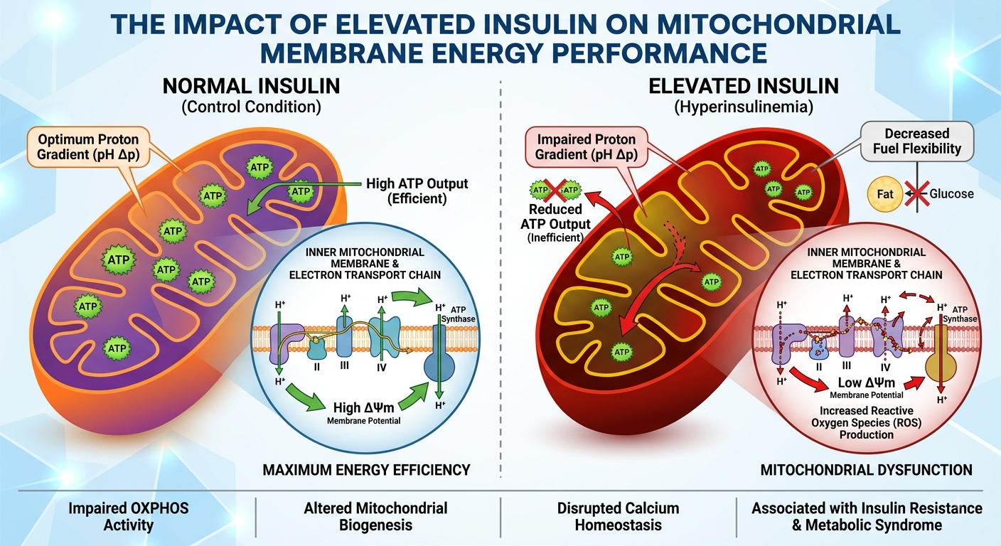
This thesis examines the relationship between insulin signaling, hyperinsulinemia, insulin resistance, mitochondrial membrane function, electron transport, proton gradients, ATP production, reactive oxygen species, and metabolic health. Particular attention is given to the distinction between normal insulin signaling and pathological metabolic states characterized by chronic insulin elevation. Insulin itself should not be regarded simply as a mitochondrial toxin. Physiological insulin signaling can support metabolic and mitochondrial processes, whereas insulin resistance and nutrient excess can produce a complex environment in which altered substrate flux, redox imbalance, lipid accumulation, inflammation, and mitochondrial dysfunction interact.
The mitochondrial inner membrane is central to this system. Electron transport through respiratory complexes I–IV establishes an electrochemical proton gradient, while ATP synthase uses the proton-motive force to convert ADP and inorganic phosphate into ATP. Understanding how insulin signaling influences the substrates and regulatory pathways feeding this machinery—and how mitochondrial dysfunction can subsequently affect insulin sensitivity—is therefore essential to understanding metabolic health.
1. Introduction
Human cellular life depends upon continuous energy conversion. Cells obtain chemical energy from nutrients and convert that energy into ATP, the principal immediately usable energy currency of the cell.
The mitochondrion performs a major part of this conversion. Carbohydrates, fats, and amino acids are metabolically processed into reducing equivalents such as NADH and FADH₂. These molecules deliver high-energy electrons to the mitochondrial respiratory chain. Electron transfer releases energy that is used to move protons across the inner mitochondrial membrane. The resulting electrochemical gradient provides the driving force for ATP synthesis.
Insulin sits upstream of many of these metabolic processes.
The simplified relationship is:
Nutrients → insulin signaling → substrate handling → mitochondrial metabolism → electron transport → proton gradient → ATP synthesis → cellular work
However, this relationship is not linear. Mitochondrial metabolism also influences insulin sensitivity. Consequently:
Insulin signaling ⇄ mitochondrial metabolism
This bidirectional relationship is particularly important in insulin resistance and metabolic disease. Research indicates that mitochondrial abnormalities and impaired insulin signaling can influence one another, although the precise causal sequence varies according to tissue, experimental model, and metabolic state.
2. The Biological Function of Insulin
Insulin is a peptide hormone produced by pancreatic β-cells. Its major physiological role is to coordinate nutrient availability with cellular metabolism.
After food intake, increasing circulating glucose stimulates insulin secretion. Insulin then acts on target tissues through the insulin receptor, initiating intracellular signaling pathways.
Major metabolic effects include:
- stimulation of glucose uptake in insulin-responsive tissues;
- promotion of glycogen synthesis;
- suppression of hepatic glucose production;
- regulation of lipid synthesis and storage;
- inhibition of excessive adipose-tissue lipolysis;
- regulation of protein metabolism;
- modulation of cellular growth and metabolic gene expression.
The classical signaling pathway can be represented as:
Insulin → insulin receptor → IRS proteins → PI3K → AKT → downstream metabolic targets
The IRS–PI3K–AKT pathway is a major mechanism through which insulin regulates nutrient and metabolic homeostasis.
3. Insulin Signaling and Cellular Energy Management
Insulin does not directly manufacture ATP.
Instead, it changes the conditions under which cells acquire, store, and metabolize energy.
For example:
Insulin signaling → increased glucose availability inside responsive cells → glycolysis → pyruvate → acetyl-CoA → TCA cycle → NADH/FADH₂ → respiratory chain → ATP
The hormone therefore functions as a metabolic coordinator.
Insulin signaling also affects lipid metabolism. Fatty acids can undergo mitochondrial β-oxidation, producing acetyl-CoA, NADH, and FADH₂. These products feed into oxidative metabolism.
Consequently, insulin can influence mitochondrial energy production indirectly by determining which substrates are available and how those substrates are processed.
4. Hyperinsulinemia
4.1 Definition
Hyperinsulinemia refers to an abnormally high concentration of insulin in the circulation.
It may occur for different reasons, including increased insulin secretion in response to insulin resistance.
A common conceptual sequence is:
Reduced insulin sensitivity → increased pancreatic insulin secretion → elevated circulating insulin → compensation for impaired insulin action
Initially, this compensation can help maintain relatively normal blood glucose.
However, persistent metabolic stress may eventually overwhelm compensatory mechanisms.
5. Hyperinsulinemia Is Not Equivalent to Mitochondrial Toxicity
An important scientific distinction is necessary.
It would be incorrect to conclude:
High insulin → directly damages mitochondria
The biology is considerably more complicated.
Normal insulin signaling can participate in maintaining mitochondrial metabolic function. Research has described relationships between insulin signaling, electron-transport-chain integrity, NAD⁺/NADH balance, SIRT1 activity, PGC-1α signaling, and mitochondrial biogenesis.
The more scientifically defensible model is:
Chronic metabolic stress + insulin resistance + altered nutrient flux + elevated insulin → altered mitochondrial regulation and redox balance
The exact contribution of hyperinsulinemia itself can vary by tissue and physiological context.
6. Insulin Resistance
Insulin resistance occurs when cells respond inadequately to insulin.
At the systemic level, this can produce compensatory insulin secretion.
The relationship can therefore become:
Insulin resistance → compensatory hyperinsulinemia → continued metabolic signaling → progressive metabolic stress
Insulin resistance is associated with obesity, metabolic syndrome, and type 2 diabetes, while its underlying molecular mechanisms involve multiple interacting processes, including inflammation, oxidative stress, endoplasmic-reticulum stress, and mitochondrial dysfunction.
7. The Mitochondrion: Cellular Energy Conversion System
A mitochondrion contains several structurally important compartments:
- outer mitochondrial membrane;
- intermembrane space;
- inner mitochondrial membrane;
- mitochondrial matrix.
The inner mitochondrial membrane is especially important for energy conversion.
It contains the respiratory-chain complexes and ATP synthase.
Unlike many biological membranes, it must maintain a tightly controlled proton gradient. This gradient stores potential energy.
The mitochondrion can therefore be conceptualized as an electrochemical energy-conversion system.
8. The Inner Mitochondrial Membrane
The inner membrane contains:
- Complex I;
- Complex II;
- Complex III;
- Complex IV;
- ATP synthase, also called Complex V;
- electron carriers;
- transport proteins;
- other proteins involved in mitochondrial metabolism.
The respiratory chain transfers electrons toward oxygen while generating the proton gradient used by ATP synthase.
The membrane therefore performs two linked functions:
electron transport
and
energy storage as an electrochemical gradient
9. Electron Transport
The respiratory chain receives electrons primarily from NADH and FADH₂.
The simplified sequence is:
NADH → Complex I → Coenzyme Q → Complex III → Cytochrome c → Complex IV → O₂
Meanwhile:
FADH₂ → Complex II → Coenzyme Q → Complex III → Cytochrome c → Complex IV → O₂
Complexes I, III, and IV contribute to proton translocation across the inner membrane.
Complex II transfers electrons into the chain but does not itself pump protons.
The respiratory chain therefore converts the energy contained in reducing equivalents into an electrochemical gradient.
10. The Proton Gradient
The electron transport chain moves protons from the mitochondrial matrix toward the intermembrane space.
This produces:
- a proton concentration difference;
- an electrical potential difference;
- an electrochemical gradient.
Together these components constitute the proton-motive force.
The mitochondrial membrane therefore behaves somewhat like an energy-storage barrier.
Energy from electron transfer is temporarily stored as electrochemical potential.
11. Mitochondrial Membrane Potential
The electrical component of the proton-motive force is commonly represented as:
Δψm
where Δψm represents the mitochondrial membrane potential.
The proton-motive force can conceptually be expressed as:
Δp = Δψ − (2.303RT/F)ΔpH
where:
- Δp = proton-motive force;
- Δψ = electrical membrane potential;
- ΔpH = pH difference;
- R = gas constant;
- T = absolute temperature;
- F = Faraday constant.
The membrane potential is therefore not simply a voltage in isolation. It is one component of a broader electrochemical energy-storage system.
12. ATP Synthase
ATP synthase converts the stored electrochemical energy of the proton gradient into chemical energy in ATP.
It consists broadly of:
- F₀, embedded in the inner mitochondrial membrane;
- F₁, projecting toward the mitochondrial matrix.
Protons flow through the membrane-associated portion, producing rotational movement that drives catalytic activity in the F₁ component.
The overall process is:
Proton gradient → proton flow → ATP synthase rotation → ADP + Pi → ATP
This molecular mechanism is supported by extensive structural and biochemical research.
13. ATP: The Cellular Energy Currency
ATP contains three phosphate groups.
Its hydrolysis can release usable free energy:
ATP + H₂O → ADP + Pi + energy
Cells use ATP to power:
- ion pumps;
- muscle contraction;
- biosynthesis;
- active transport;
- cellular signaling;
- protein turnover;
- membrane maintenance;
- electrical activity.
Mitochondrial oxidative phosphorylation therefore provides a major energy supply for cellular work.
14. Insulin and Mitochondrial Substrate Availability
Insulin influences the availability and utilization of metabolic substrates.
Glucose can undergo:
Glucose → glycolysis → pyruvate → acetyl-CoA → TCA cycle
Fatty acids can undergo:
Fatty acid → β-oxidation → acetyl-CoA + NADH + FADH₂
These pathways generate the reducing equivalents required for oxidative phosphorylation.
Consequently, insulin can alter mitochondrial energy metabolism through upstream regulation of nutrient utilization.
15. NADH and FADH₂
NADH and FADH₂ are electron carriers.
They transfer electrons generated through metabolic pathways to the mitochondrial respiratory chain.
The simplified relationship is:
Nutrient oxidation → NADH/FADH₂ → electron transport → proton pumping → ATP
The NAD⁺/NADH ratio is also an important indicator of cellular redox state.
Alterations in this ratio can influence metabolic enzymes and signaling pathways.
Research has connected insulin signaling with maintenance of NAD⁺/NADH balance and pathways involving SIRT1 and PGC-1α.
16. Insulin, SIRT1, and PGC-1α
SIRT1 is a NAD⁺-dependent deacetylase involved in metabolic regulation.
PGC-1α is a major regulator of mitochondrial biogenesis and oxidative metabolism.
A simplified pathway is:
NAD⁺ availability → SIRT1 activity → PGC-1α regulation → mitochondrial gene programs
Insulin resistance can disrupt components of this network.
This provides one possible molecular bridge between impaired insulin signaling and mitochondrial adaptation.
17. Mitochondrial Biogenesis
Mitochondrial biogenesis refers to the generation and expansion of mitochondrial components.
It involves:
- nuclear gene expression;
- mitochondrial gene expression;
- protein synthesis;
- mitochondrial protein import;
- respiratory-complex assembly;
- membrane formation;
- mitochondrial division.
Mitochondrial quantity, quality, turnover, and functional capacity are therefore distinct concepts.
A greater number of mitochondria does not automatically mean better mitochondrial performance.
18. Oxidative Stress
Electron transport is not perfectly efficient.
A small proportion of electrons can participate in reactions that generate reactive oxygen species (ROS).
Important ROS include:
- superoxide;
- hydrogen peroxide;
- hydroxyl radicals under appropriate chemical conditions.
ROS are not universally harmful.
At controlled concentrations they participate in signaling.
Excessive or poorly controlled ROS can, however, damage:
- lipids;
- proteins;
- DNA;
- mitochondrial components;
- signaling proteins.
Research indicates that ROS can both participate in normal insulin signaling and contribute to insulin resistance when oxidative stress becomes excessive.
19. The Mitochondrial Membrane and Oxidative Damage
The inner mitochondrial membrane is particularly important because it contains the molecular machinery responsible for oxidative phosphorylation.
Oxidative damage affecting membrane lipids or respiratory proteins can alter:
- electron transfer;
- proton pumping;
- membrane potential;
- proton permeability;
- ATP production;
- respiratory efficiency.
The result can be reduced energetic efficiency or altered redox signaling.
20. Proton Leak and Uncoupling
Not all protons returning to the mitochondrial matrix necessarily pass through ATP synthase.
Some can return through alternative pathways.
This is called proton leak.
A simplified energy model is:
Electron transport → proton gradient → ATP synthase → ATP
versus:
Electron transport → proton gradient → proton leak → heat
Controlled uncoupling has physiological roles, but excessive uncoupling can reduce the amount of ATP produced per unit of substrate oxidation.
Research in insulin-resistant and insulin-deficient states has reported altered mitochondrial coupling and increased ROS production.
21. ATP Production Efficiency
Mitochondrial performance should not be measured simply by asking:
“How much oxygen does the mitochondrion consume?”
A complete analysis should consider:
- oxygen consumption;
- ATP production;
- coupling efficiency;
- respiratory capacity;
- membrane potential;
- proton leak;
- ROS production;
- substrate oxidation;
- mitochondrial density;
- mitochondrial dynamics.
A mitochondrion can maintain substantial oxygen consumption while producing ATP inefficiently if coupling is impaired.
22. Hyperinsulinemia, Nutrient Excess, and Mitochondrial Stress
Chronic metabolic excess can create increased substrate delivery to mitochondria.
Potential consequences include:
Excess nutrient availability → increased reducing equivalents → increased electron pressure → altered electron transport → increased ROS under some conditions → oxidative stress
This does not mean that every episode of high insulin produces mitochondrial damage.
Rather, persistent metabolic overload can change the environment in which mitochondrial metabolism operates.
23. Lipid Overload
Fatty acids are important mitochondrial fuels.
However, excessive lipid availability can produce metabolic intermediates that interfere with insulin signaling and cellular homeostasis.
The relationship can be represented as:
Excess fatty-acid availability → altered lipid intermediates → impaired insulin signaling → altered substrate metabolism → mitochondrial stress
The liver and skeletal muscle are particularly important tissues in this relationship.
24. Skeletal Muscle
Skeletal muscle is one of the largest sites of insulin-stimulated glucose disposal.
Its mitochondria are therefore central to whole-body metabolic health.
Insulin resistance in skeletal muscle can affect:
- glucose uptake;
- glycogen synthesis;
- fatty-acid oxidation;
- mitochondrial substrate utilization;
- ATP production;
- metabolic flexibility.
Human research demonstrates associations between mitochondrial characteristics and insulin sensitivity, although the findings are not universally consistent.
25. Liver
The liver coordinates systemic metabolism.
It regulates:
- glucose production;
- glycogen metabolism;
- fatty-acid synthesis;
- fatty-acid oxidation;
- lipoprotein metabolism;
- ketone-body production.
Insulin resistance in the liver can disrupt these processes and contribute to metabolic disease.
Mitochondrial dysfunction may consequently become part of a broader cycle involving altered lipid and glucose metabolism.
26. Adipose Tissue
Adipose tissue is an endocrine and metabolic organ.
Insulin normally suppresses excessive lipolysis.
When insulin signaling becomes impaired, increased fatty-acid release from adipose tissue can contribute to lipid delivery to other organs.
This can further influence:
liver metabolism → skeletal-muscle metabolism → insulin sensitivity → systemic metabolic health
27. The Mitochondria–Insulin Resistance Feedback Loop
A useful conceptual model is:
Insulin resistance
↓
Compensatory hyperinsulinemia
↓
Altered nutrient handling
↓
Increased metabolic stress
↓
Mitochondrial redox and energetic alterations
↓
ROS and cellular stress
↓
Further impairment of insulin-signaling pathways
↓
Greater insulin resistance
This represents a conceptual feedback system rather than a universal sequence occurring identically in every person or tissue.
28. Cause or Consequence?
One of the most important scientific questions is:
Does mitochondrial dysfunction cause insulin resistance, or does insulin resistance cause mitochondrial dysfunction?
Current evidence does not support a simple universal answer.
Studies have reported:
- mitochondrial abnormalities preceding metabolic dysfunction;
- mitochondrial changes appearing after metabolic stress;
- tissue-specific effects;
- differences between mitochondrial quantity and mitochondrial function;
- differences between animal models and humans.
Reviews therefore describe the relationship as complex and potentially bidirectional.
29. The Role of Reactive Oxygen Species
ROS can be represented as both:
Signaling molecules
Moderate ROS levels can participate in cellular signaling, including insulin signaling.
Stress mediators
Persistent excessive ROS can modify proteins and lipids and interfere with signaling networks.
Therefore:
ROS ≠ automatically harmful
Instead:
ROS balance = physiological signaling + antioxidant control + metabolic context
The problem arises when ROS production exceeds the capacity of cellular systems to regulate and repair oxidative damage.
30. Antioxidant Defense
Cells possess antioxidant systems including:
- superoxide dismutases;
- catalase;
- glutathione systems;
- peroxiredoxins;
- thioredoxin systems.
These systems maintain redox balance.
Mitochondrial health therefore depends not merely on minimizing ROS but on maintaining an appropriate relationship between:
ROS production ↔ ROS signaling ↔ antioxidant defense ↔ repair
31. Mitochondrial Quality Control
Mitochondria are continuously remodeled.
Important processes include:
Fusion
Mitochondria combine their contents.
Fission
Mitochondria divide.
Mitophagy
Damaged mitochondria can be selectively removed through autophagic mechanisms.
Biogenesis
New mitochondrial components are produced.
Together these processes create mitochondrial quality control.
Metabolic disease can disrupt mitochondrial dynamics and turnover. Contemporary research continues to investigate how these changes interact with insulin resistance.
32. Mitochondrial DNA
Mitochondria contain their own genome.
Mitochondrial DNA encodes a subset of proteins required for oxidative phosphorylation.
Because mitochondria are exposed to intense metabolic activity and ROS generation, mitochondrial genetic integrity is important for long-term respiratory function.
Damage to mitochondrial DNA can potentially affect respiratory-chain performance and cellular metabolism.
33. Mitochondrial Respiratory Supercomplexes
Respiratory complexes do not necessarily operate as completely isolated entities.
They can form higher-order assemblies known as respiratory supercomplexes.
These structures may influence:
- electron transfer;
- respiratory efficiency;
- complex stability;
- ROS production.
Modern mitochondrial biology therefore increasingly considers the respiratory chain as an organized membrane system rather than merely five independent enzyme complexes.
34. Energy Performance as a Systems Property
Mitochondrial energy performance cannot be reduced to one variable.
A comprehensive model is:
Energy performance = substrate oxidation + electron transfer + proton pumping + membrane integrity + proton-motive force + ATP synthase activity + nucleotide exchange + redox control
Failure at any stage can influence the final ATP output.
35. Insulin and Metabolic Flexibility
Healthy metabolism requires the ability to switch between fuels.
For example:
Fed state → greater glucose utilization
and
fasted/exercise state → greater reliance on fatty-acid oxidation
Insulin is one of the principal regulators of this transition.
Insulin resistance can reduce metabolic flexibility, causing tissues to respond less appropriately to changes in nutrient availability.
36. Exercise and Mitochondrial Adaptation
Physical activity places controlled energetic demands on skeletal muscle.
Repeated exercise can stimulate mitochondrial adaptations involving:
- mitochondrial biogenesis;
- oxidative capacity;
- substrate utilization;
- antioxidant defenses;
- metabolic flexibility.
Exercise therefore provides an important physiological model for studying how energy demand can influence mitochondrial capacity and insulin sensitivity.
37. Nutrition and Mitochondrial Metabolism
Mitochondria respond continuously to nutrient availability.
Major fuel classes include:
Carbohydrates → glucose → pyruvate → acetyl-CoA
Fats → fatty acids → β-oxidation → acetyl-CoA
Proteins → amino acids → metabolic intermediates
The metabolic system must coordinate these inputs with cellular energy demand.
Persistent nutrient excess can produce a different metabolic environment from temporary nutrient availability.
38. Experimental Measurement of Mitochondrial Function
Scientific studies can assess mitochondrial performance through several approaches.
Important measurements include:
Oxygen consumption
Measures respiratory activity.
Respiratory capacity
Examines the maximum capacity of the respiratory system under defined experimental conditions.
ATP production
Assesses cellular energy output.
Membrane potential
Examines the electrical component of the proton-motive force.
ROS production
Measures oxidative signaling or oxidative stress.
Respiratory-chain activity
Examines individual complexes or integrated respiratory function.
Mitochondrial content
Assesses mitochondrial abundance.
No single measurement completely defines mitochondrial health.
39. A Quantitative View of Oxidative Phosphorylation
The core energy relationship can be summarized as:
NADH/FADH₂ oxidation
↓
Electron transport
↓
Proton translocation
↓
Δp
↓
ATP synthase
↓
ATP
The proton-motive force contains both electrical and chemical components:
Δp = membrane-potential component + pH-gradient component
ATP synthesis is therefore fundamentally an electrochemical process.
40. A Systems Model of Insulin–Mitochondrial Interaction
The entire system can be summarized as:
Insulin
↓
Insulin receptor
↓
IRS–PI3K–AKT signaling
↓
Glucose and lipid regulation
↓
Substrate availability
↓
Glycolysis / β-oxidation
↓
TCA cycle
↓
NADH + FADH₂
↓
Electron transport chain
↓
Proton pumping
↓
Mitochondrial membrane potential
↓
ATP synthase
↓
ATP
↓
Cellular work
At the same time:
Mitochondrial dysfunction
↓
Altered ROS / redox balance
↓
Modification of insulin-signaling components
↓
Insulin resistance
This establishes the central bidirectional relationship of the thesis.
41. Metabolic Health
Metabolic health depends upon coordination among:
- insulin sensitivity;
- glucose regulation;
- lipid metabolism;
- mitochondrial function;
- redox balance;
- inflammation;
- energy expenditure;
- tissue communication.
Mitochondrial health is therefore one component of metabolic health rather than an isolated phenomenon.
42. The Central Scientific Question
The fundamental question is not simply:
“Does high insulin damage mitochondria?”
A better scientific question is:
How do insulin concentration, insulin signaling, nutrient availability, insulin resistance, mitochondrial substrate oxidation, membrane energetics, redox balance, and cellular energy demand interact over time?
This framing avoids reducing a complex biological network to a single hormone-toxicity relationship.
43. Integrated Pathophysiological Model
A comprehensive model can be represented as:
Nutrient excess / metabolic stress
↓
Insulin resistance
↓
Compensatory hyperinsulinemia
↓
Altered glucose and lipid handling
↓
Altered mitochondrial substrate flux
↓
Changes in NADH/FADH₂ supply
↓
Changes in respiratory-chain activity
↓
Changes in proton gradient
↓
Changes in membrane potential and coupling
↓
Altered ATP/ROS balance
↓
Cellular oxidative and metabolic stress
↓
Further impairment of metabolic signaling
This is a systems-level model, and the magnitude and direction of each step depend on tissue and physiological context.
44. Important Scientific Qualifications
Several conclusions should be avoided:
Incorrect simplification 1
“Insulin is bad for mitochondria.”
Insulin is physiologically necessary and participates in normal metabolic regulation.
Incorrect simplification 2
“High insulin automatically means mitochondrial damage.”
Hyperinsulinemia can occur in different metabolic contexts, and mitochondrial effects cannot be inferred from insulin concentration alone.
Incorrect simplification 3
“Mitochondrial dysfunction always causes insulin resistance.”
Evidence indicates a complex and potentially bidirectional relationship.
Incorrect simplification 4
“All ROS are harmful.”
Controlled ROS production has physiological signaling functions.
45. Major Research Questions
Future research should investigate:
- Which mitochondrial defects occur first during development of insulin resistance?
- How does chronic hyperinsulinemia independently affect mitochondrial function?
- How do glucose and fatty-acid overload interact at the mitochondrial level?
- How does mitochondrial membrane composition affect insulin sensitivity?
- How does mitochondrial membrane potential change during insulin resistance?
- How does proton leak alter whole-cell energy efficiency?
- Which respiratory-chain complexes are most vulnerable to metabolic stress?
- How do mitochondrial supercomplexes respond to metabolic disease?
- How does mitochondrial ROS modify insulin signaling?
- How does insulin signaling regulate mitochondrial biogenesis?
- How do skeletal muscle and liver mitochondria differ?
- What role does mitophagy play in maintaining insulin sensitivity?
- Can mitochondrial dysfunction be reversed?
- Which mitochondrial biomarkers best predict metabolic disease?
- How should mitochondrial function be measured in living humans?
46. Conclusion
Insulin and mitochondria belong to the same integrated metabolic system.
Insulin regulates the cellular handling of nutrients, while mitochondria convert the chemical energy contained in those nutrients into ATP. The mitochondrial respiratory chain transfers electrons and uses the released energy to establish a proton-motive force across the inner mitochondrial membrane. ATP synthase then converts that electrochemical energy into ATP.
The relationship becomes more complex in insulin resistance and hyperinsulinemia. Chronic metabolic stress can alter substrate flux, redox balance, mitochondrial dynamics, oxidative phosphorylation, ROS production, and insulin signaling. At the same time, mitochondrial dysfunction may itself contribute to impaired insulin sensitivity. Modern evidence therefore supports a bidirectional, tissue-specific metabolic relationship rather than a simple model in which elevated insulin directly damages mitochondrial membranes.
The mitochondrial inner membrane is the critical energetic interface connecting these processes. It contains the molecular machinery that transforms electron flow into a proton gradient and subsequently transforms that gradient into ATP.
The central principle can therefore be summarized as:
Insulin signaling regulates nutrient availability and metabolic flux → nutrient oxidation generates NADH and FADH₂ → the respiratory chain converts electron energy into a proton gradient → the mitochondrial membrane stores electrochemical potential → ATP synthase converts that potential into ATP → ATP powers cellular life.
When metabolic regulation becomes chronically disturbed, this finely coordinated system can develop abnormalities involving insulin signaling, substrate overload, oxidative stress, mitochondrial coupling, membrane potential, and ATP production.
Understanding this network provides a more scientifically accurate picture of metabolic health than focusing on insulin concentration alone.
Core Scientific Framework
INSULIN SIGNALING
↓
GLUCOSE + LIPID METABOLISM
↓
GLYCOLYSIS + β-OXIDATION + TCA CYCLE
↓
NADH + FADH₂
↓
ELECTRON TRANSPORT CHAIN
↓
PROTON TRANSLOCATION
↓
MITOCHONDRIAL PROTON-MOTIVE FORCE
↓
MEMBRANE POTENTIAL + pH GRADIENT
↓
ATP SYNTHASE
↓
ATP
↓
CELLULAR ENERGY
↕
ROS / REDOX BALANCE
↕
INSULIN SENSITIVITY
This bidirectional architecture is the anatomy of the insulin–mitochondrial energy relationship and provides the central scientific framework for understanding hyperinsulinemia, mitochondrial bioenergetics, oxidative stress, insulin resistance, and metabolic health.





Be First to Comment